
1. Architectural Overview: Why Global Procurement Is Transitioning to 3D Skin Analysis Machines
Across the global cosmetic, pharmaceutical, and medical aesthetics landscapes, traditional 2D optical photography is rapidly proving insufficient for rigorous claim substantiation and high-converting patient consultations. Standard planar imaging, while useful for macroscopic color observation, suffers from critical physical constraints: light reflection variability, operator angle inconsistency, micro-shadowing distortion, and an absolute inability to extract true quantitative depth ($z$-axis depth measurements in micrometers).
A clinical-grade 3D skin analysis machine solves these empirical deficits by capturing surface topography along with spectral absorption data. By utilizing multi-directional, multi-wavelength LED illumination, modern 3D skin analysis hardware constructs an exact mathematical mesh of the cutaneous surface. This enables researchers and clinicians to measure parameters that were previously unquantifiable without invasive biopsies or cumbersome silicone replicas—such as wrinkle volume ($mm^3$), skin roughness metrics ($Ra$ and $Rz$), fine line indentation indices, pore depth, epidermal melanin distribution, and dermal hemoglobin concentrations.
Search Intent Insight: Information Gain for B2B Buyers
Most commercial buying guides focus solely on marketing aesthetics. From an engineering and clinical trial standpoint, a true 3D skin analysis machine must be evaluated on three non-negotiable physical dimensions: illumination uniformity, photometric reconstruction accuracy, and spectral unmixing capabilities. Without calibrated multispectral LEDs and mathematical diffuse reflection modeling, system outputs are merely color-manipulated 2D images rather than validated 3D dermatological datasets.
Quantifiable Physics Over Visual Guesswork
Clinical efficacy demands repeatable data. Whether validating a novel hyaluronic acid topical formulation in a double-blind CRO study or demonstrating the 90-day resurfacing outcome of a fractional $CO_2$ laser treatment, quantitative parameters serve as the universal language of proof.
Modern 3D skin imaging systems eliminate subjectivity. By acquiring skin topography at sub-millimeter resolutions, clinical trial managers receive standard-deviation-backed metrics suitable for regulatory submissions, peer-reviewed medical publications, and high-trust patient education.
2. Technical Deep-Dive: Product Capabilities of the Antera 3D® System
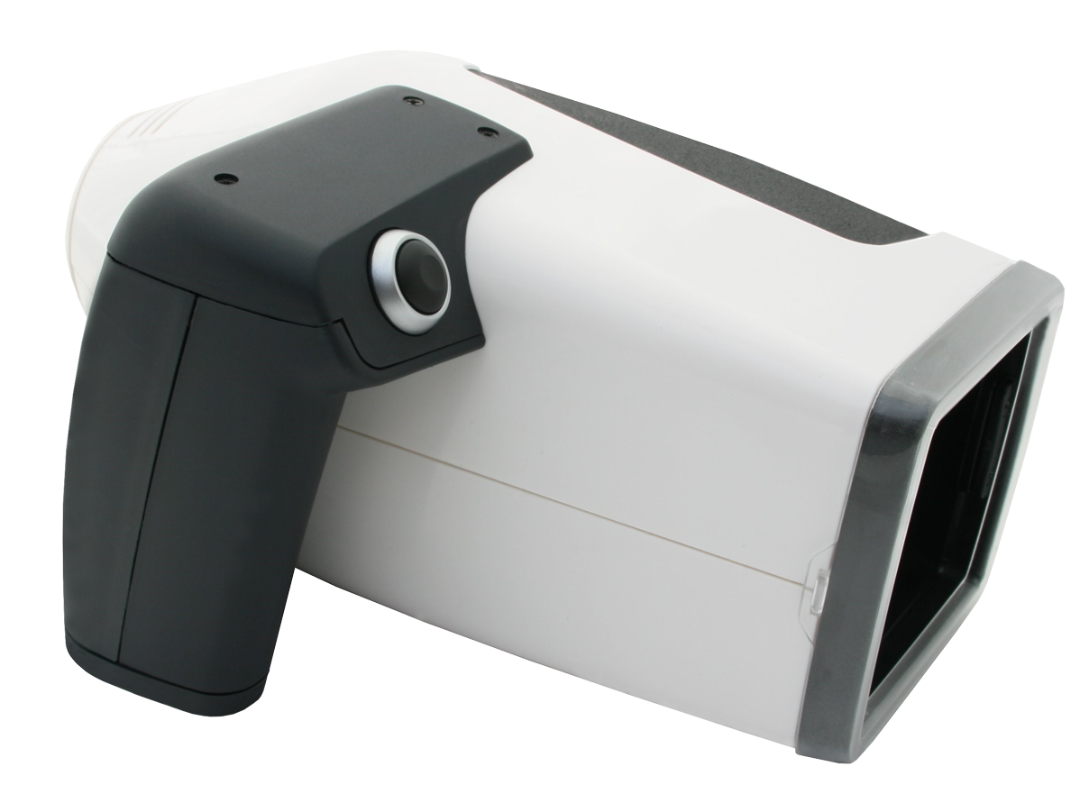
Developed by Miravex Limited (headquartered at 11 St. Stephen's Green, Dublin, Ireland), the Antera 3D® skin analysis machine represents the gold standard in portable, high-resolution dermatological imaging. Derived from advanced optical research at Trinity College Dublin, Antera 3D® utilizes patented photometric stereo imaging to transform multi-angle lighting data into precise 3D elevation maps.
2.1 Core Optical Principles: Photometric Stereo & Multispectral Analysis
Unlike conventional 3D scanners that rely on structured light projection (which can blur at microscopic cutaneous scales) or passive stereovision (which struggles with textureless skin surfaces), the Antera 3D® machine utilizes Photometric Stereo Imaging. The optical unit features arrayed LEDs emitting light from multiple known spatial angles and distinct spectral wavelengths ranging from the visible spectrum to the near-infrared.
- Topographical Mesh Generation: By capturing surface reflectivity variations as lighting angles shift milliseconds apart, the software computes surface normal vectors for every pixel, reconstructing skin micro-relief with precision down to 1 micrometer ($\mu m$).
- Beer-Lambert Spectral Unmixing: Utilizing light absorption properties at varied wavelengths, the Antera 3D® software isolates skin chromophores. It differentiates epidermal melanin (hyperpigmentation, sun damage, age spots) from dermal hemoglobin (erythema, rosacea, vascular telangiectasias, inflammation).
| Measurement Parameter | Technical Metric / Unit | Clinical & Commercial Application |
|---|---|---|
| Wrinkle Depth & Volume | Anti-aging cream clinical trials, neurotoxin & dermal filler efficacy documentation. | |
| Skin Texture & Roughness | Exfoliation studies, microdermabrasion tracking, skin barrier repair evaluation. | |
| Melanin Concentration | Hyperpigmentation treatment efficacy, whitening cream testing, melasma therapy monitoring. | |
| Hemoglobin & Redness | Rosacea clinical management, post-laser inflammation reduction, IPL treatment tracking. | |
| Pore Characteristics | Astringent formulation testing, sebum control evaluation, acne treatment follow-ups. | |
| 3D Color Topography | Pre/post-procedure visual proof for plastic surgery and aesthetic consultations. |
3. Global Procurement Trends: Future-Proofing 3D Skin Analysis Investment
As healthcare technology buyers, contract research organizations (CROs), and medical aesthetic directors evaluate 3D skin analysis machines for 2025 and beyond, several structural market shifts are redefining hardware and software requirements:
Trend 1: Decentralized Clinical Trials (DCTs) & Portable Ergonomics
Massive, rigid full-face imaging booths are increasingly viewed as space-inefficient bottlenecks in modern clinical workflow environments. Procurement preferences have shifted heavily toward ergonomic, portable hand-held 3D skin analyzers. Portable instruments like the Antera 3D® allow investigators to analyze any anatomical region—including cheeks, periorbital zones, neck, décolletage, hands, and arms—with equal spatial accuracy, enabling multi-center trial consistency.
Trend 2: AI-Driven Predictive Dermatological Modeling
Artificial intelligence integration has moved from a novelty feature to a core analytical requirement. Procurement teams now prioritize skin imaging platforms capable of automated ROI (Region of Interest) matching. When subjects return for 30-, 60-, or 90-day follow-ups, advanced image-registration algorithms align past and present scans pixel-by-pixel, eliminating operator position variability and ensuring strict mathematical comparability.
Trend 3: Stringent Regulatory & Advertising Efficacy Requirements
Regulatory bodies globally—including the US FTC, European Medicines Agency (EMA), and China National Medical Products Administration (NMPA)—are enforcing strict evidentiary standards for skincare marketing claims. Soft claims like "skin appears 80% smoother" must now be backed by objective physical metrics: "statistically significant reduction of 24.3% in wrinkle volume ($p < 0.001$) measured via 3D photometric stereo imaging."
Trend 4: Integration with Electronic Health Records (EHR) & Enterprise CDMS
Scalability requires seamless data export. Enterprise buyers demand skin analysis software capable of exporting raw vector arrays, high-resolution CSV datasets, DICOM-compatible files, and customized PDF clinical reports. This ensures seamless sync with Clinical Data Management Systems (CDMS) and aesthetic practice management software.
Clinical Video Demonstration & Workflow Integration
Watch how the Antera 3D® software captures, reconstructs, and quantifies complex skin lesions and wrinkle topography in real time. The seamless user interface enables rapid acquisition under 2 seconds per scan.
High-speed acquisition minimizes motion artifacts, ensuring high compliance even during large-scale clinical screening trials involving hundreds of subjects daily.
4. Technology Evolution Roadmap: The Future of 3D Skin Analysis (2025–2030)
Looking toward the next decade of dermatological science, the technological matrix underpinning 3D skin analysis machines will evolve across four core domains:
- Hyper-Spectral Deep Tissue Penetration: Future iterations will expand beyond standard RGB and near-infrared spectra into mid-infrared wavelengths, providing non-invasive volumetric optical coherence tomography (OCT) capabilities to map dermal collagen bundle alignment without biopsies.
- Real-Time Generative Simulation of Biological Aging: Leveraging generative AI models trained on millions of longitudinal 3D skin scans, skin analysis machines will project accurate 5-year and 10-year skin degradation trajectories based on current solar damage, elastin loss, and vascular inflammation levels.
- Automated Biomarker Cross-Correlation: Algorithmic software will correlate surface topography variations with biological micro-inflammation markers, providing holistic insights that connect topical skin barrier dysfunction to systemic health metrics.
- Sub-Micrometer Contact Topography: Advancements in optical sensor density will push 3D resolution into sub-micron territory, enabling the optical visualization of individual cell clusters, hair follicle sebum impaction depth, and micro-capillary loop structures.
5. Enterprise Advantages of Partnering with Miravex Limited
When acquiring medical-grade skin analysis equipment, selecting the right technology partner is as vital as hardware specifications. Miravex Limited stands out as a world leader in quantitative skin analysis technology:
- 15+ Years of Scientific Innovation: Founded as a spin-out from Trinity College Dublin's prestigious Physics Department, Miravex brings deep optical physics rigor to commercial dermatological instrumentation.
- Validated by 500+ Peer-Reviewed Publications: Antera 3D® is not an unverified commercial scanner; it is a clinical benchmark referenced in over 500 scientific papers published in top-tier journals such as the Journal of Cosmetic Dermatology, Dermatologic Surgery, and Skin Research and Technology.
- Global Presence in 40+ Countries: Trusted by global cosmetic conglomerates (L'Oréal, Estée Lauder, Beiersdorf, Procter & Gamble), tier-1 CROs (Eurofins, SGS, Dermscan), and top aesthetic practices across Europe, North America, Asia-Pacific, and the Middle East.
- Strict Compliance & Data Integrity: Designed for clinical trial integrity, Antera 3D® software offers audit-trail logging, GDPR/HIPAA compliant data architecture, and reproducible calibration routines that guarantee zero drift across multi-year multi-center studies.
6. Comprehensive B2B Procurement FAQ: Answering Key Questions
To assist procurement committees, clinical managers, and practice owners, we have compiled detailed answers to the most frequent technical and commercial queries submitted to our engineering team:
Q1: What is the primary technical difference between a hand-held 3D skin analysis machine like Antera 3D® and a 2D full-face imaging box?
Traditional 2D full-face imaging boxes capture planar surface photographs using fixed diffuse lighting. While they offer macro-visual comparison of facial symmetry, they cannot measure vertical depth, volumetric changes, or surface roughness ($Ra/Rz$) because 2D pixels lack spatial $z$-coordinate data.
In contrast, a hand-held 3D skin analysis machine utilizing photometric stereo optics captures multi-directional illuminated images at microscopic resolutions. It computes true spatial 3D surface topography, allowing quantitative depth measurements down to 1 micrometer. Furthermore, hand-held units allow target-specific imaging anywhere on the human body (e.g., neck, hands, body contours), whereas full-face booths are restricted strictly to facial areas.
Q2: How does photometric stereo reconstruction calculate true 3D skin roughness (Ra/Rz) and wrinkle volume?
Photometric stereo relies on capturing multiple images of the exact same skin surface area illuminated sequentially from different known angular light sources. Because skin micro-relief casts microscopic shadows based on light elevation and azimuth, the software calculates the local surface normal (inclination) for every image pixel.
By integrating these surface normals across the image plane, the software generates a continuous 3D elevation map. Wrinkle volume ($mm^3$) is calculated by establishing a virtual reference surface across the crease margin and integrating the spatial volume below that surface. Roughness parameters ($Ra$, the arithmetic average of profile heights, and $Rz$, the mean peak-to-valley height) are calculated according to ISO 4287 industrial surface texture standards.
Q3: Can a 3D skin analysis machine accurately differentiate epidermal melanin from dermal hemoglobin across high Fitzpatrick skin phototypes?
Yes, provided the system utilizes calibrated multispectral illumination and light propagation algorithms. The Antera 3D® system uses targeted LED wavelengths across the visible spectrum where hemoglobin and melanin display distinct spectral absorption characteristics (based on the Beer-Lambert law of extinction coefficients).
By mathematically unmixing spectral absorption maps, the software separates epidermal melanin (concentrated in the basal layer) from deeper dermal hemoglobin (vascular networks). This spectral unmixing remains robust and accurate across Fitzpatrick Skin Types I through VI, avoiding the misclassification common in RGB-based camera filtration systems.
Q4: What is the typical return on investment (ROI) timeline for an aesthetic medical clinic acquiring an Antera 3D® system?
For aesthetic medical practices, plastic surgery centers, and medical spas, the Antera 3D® machine yields a dual revenue impact: boosting treatment conversion rates and increasing patient retention. Empirical practice data indicates an average ROI realization within 3 to 6 months of deployment.
During consultations, presenting patients with objective 3D baseline metrics (e.g., deep-layer sun damage, early fine line micro-relief, un-occluded redness) dramatically increases consultation-to-procedure conversion rates by up to 35%. Furthermore, providing objective visual and quantitative 3D before-and-after proof at post-treatment follow-ups increases package renewals and preventative treatment adherence.
Q5: How do cosmetic contract research organizations (CROs) use 3D skin analysis data to substantiate anti-aging efficacy claims?
Cosmetic CROs and R&D laboratories require statistically significant, objective endpoint data to substantiate claims such as "reduces wrinkle volume by 20% in 28 days" or "clinically proven to target deep hyperpigmentation."
Using Antera 3D®, study investigators capture baseline measurements ($T_0$) for trial participants. Following topical application over designated study periods ($T_{14}$, $T_{28}$, $T_{60}$), the software automatically aligns follow-up scans using image-registration algorithms. The system then outputs raw numerical data (wrinkle depth, texture parameters, melanin intensity indices) suitable for ANOVA and paired $t$-test statistical validation required by global consumer protection regulators.
Q6: What calibration protocols and maintenance routines are required to maintain clinical repeatability over long-term multi-center studies?
To ensure multi-center clinical consistency, the Antera 3D® machine utilizes an automated quick-calibration target system. Prior to study sessions, operators perform a 10-second optical target verification using a certified reference block supplied with the device.
The software automatically verifies LED luminous intensity, spectral balance, and camera sensor response, applying internal correction factors if ambient thermal shifts occur. Because the device has no moving optical parts or complex mechanical lasers, hardware drift is negligible, ensuring identical data reproducibility across international multi-center trials over several years.
Q7: How does the software handle repositioning artifacts during multi-month longitudinal clinical studies?
Skin repositioning errors are a primary source of measurement variance in clinical dermatological trials. Antera 3D® solves this through proprietary 2D/3D cross-correlation image registration.
When loading a follow-up scan, the software identifies natural anatomical landmarks (moles, micro-vascular patterns, pore clusters) and mathematically locks the follow-up Region of Interest (ROI) to the baseline ROI. Even if the camera is applied at a slightly shifted angle or position, the software corrects the alignment matrix before running quantitative comparisons, eliminating manual cropping bias.
Q8: What software integration capabilities (API / Data Export formats) exist for enterprise Clinical Data Management Systems (CDMS)?
The Antera 3D® software platform is engineered for seamless enterprise data integration. All quantitative metrics (wrinkle volumes, roughness values, chromophore indexes, surface spatial parameters) can be exported directly into structured CSV, Microsoft Excel, and SQL-compatible database formats.
Additionally, high-resolution 3D texture maps and spectral overlays can be exported as standard image files (PNG, TIFF) and vector PDFs. For large clinical trial sponsors, customized API data bridges can be deployed to auto-populate Electronic Case Report Forms (eCRFs) within major CDMS environments while maintaining strict 21 CFR Part 11 audit compliance.
7. Procurement Decision Matrix: Evaluating Your 3D Skin Analysis Investment
Selecting the optimal 3D skin analysis machine requires evaluating your institution's specific clinical, research, and financial objectives. Use the decision matrix below to align hardware features with operational requirements:
| Evaluation Criteria | Cosmetic R&D & CRO Requirements | Aesthetic Clinic & Plastic Surgery Requirements |
|---|---|---|
| Primary Objective | Statistical claim substantiation, publication-grade repeatability. | Patient conversion, visual education, procedure validation. |
| Key Metrics Needed | $Ra, Rz$ roughness, wrinkle volume ($mm^3$), absolute melanin indices. | Before/after 3D comparisons, erythema maps, pore reduction visualizer. |
| Anatomical Versatility | Critical: must scan facial, bodily, and off-face test sites. | High: facial focus, neck/chest, hands, post-liposuction sites. |
| Data Portability | CSV export, statistical software integration, audit trails. | Custom branded PDF reports, quick consultation export. |
| Recommended System | Antera 3D® CS (Clinical Suite) | Antera 3D® Standard / Aesthetic Edition |
8. Request Official Technical Specifications & Direct Quotation
Investing in a medical-grade 3D skin analysis machine is a strategic operational decision that elevates research validity and clinical revenue. As the original manufacturer of Antera 3D®, Miravex Limited provides direct global shipping, comprehensive clinical training, multi-year warranty coverage, and dedicated technical support.
Click below to connect immediately with our sales engineering team for technical datasheets, customized clinical ROI estimates, or direct procurement pricing.
Get In Touch
Contact Miravex
Speak with our team of skin science specialists. Whether you are a researcher, clinician or procurement professional, we are here to support your requirements.
Ready to Upgrade Your Skin Analysis Technology?
Discover how Antera 3D® can transform your skin research, clinical practice or product development. Our specialists are available to provide tailored guidance and demonstrations.
Inquire Now