B2B Clinical Procurement & Technical Analysis Next-Generation Clinical Dermatology Skin Scanner:
Quantitative 3D Imaging & Strategic Global Procurement Guide
An authoritative evaluation for dermatologists, pharmaceutical contract research organizations (CROs), cosmetic R&D directors, and hospital procurement teams evaluating precision multispectral 3D skin analysis equipment.
The Paradigm Shift in Dermatology: From Subjective Assessment to Quantitative 3D Optical Metrology
In contemporary clinical dermatology, aesthetic medicine, and pharmaceutical trial research, visual inspections and qualitative grading scales (such as the Fitzpatrick scale or subjective visual erythema scoring) no longer satisfy the rigorous demands of evidence-based clinical practice and regulatory compliance. Global procurement directors, clinical trial leaders, and cosmetic formulators are increasingly transitioning toward advanced clinical dermatology skin scanner systems that provide reproducible, objective, and mathematical quantification of the skin's surface and subsurface micro-structure.
Legacy 2D photography systems often suffer from significant lighting inconsistencies, parallax errors, specular reflection distortion, and an inability to isolate optical depth. By contrast, modern clinical dermatology skin scanners leverage advanced optical physics—specifically photometric stereo imaging combined with multi-wavelength light-emitting diodes (LEDs)—to capture high-resolution dermal topographies and sub-surface spectral optical density measurements in real time.
Key Information Gain for Clinical Procurement Officers:
A clinical-grade skin scanner must measure both geometric topography (wrinkle depth, fine line volume, pore depression) and chromophore spatial concentration (melanin index and hemoglobin index) without relying on artificial software filters or destructive ambient light exposure. True quantitative accuracy requires calibrated spatial illumination and contact optical standardization.
By capturing multiple images illuminated from varying directional vectors, the scanner reconstructs the 3D surface matrix with sub-micron elevation accuracy. This enables clinical researchers to measure variations in skin roughness ($R_a$, $R_z$ values), epidermal volume loss, vascular dilation, and localized hyper-pigmentation with unmatched inter-test reliability and intra-operator repeatability ($CV < 2\%$).
Recommended Industry Standard: Antera 3D® System by Miravex Limited
When evaluating commercial and clinical options in the global market, the Antera 3D® developed by Miravex Limited (Dublin, Ireland) stands out as the benchmark clinical dermatology skin scanner for medical institutions, cosmetic manufacturers, and Contract Research Organizations (CROs).
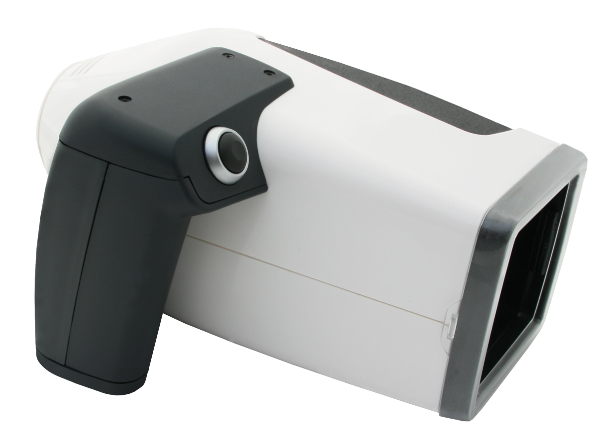
Figure 1: Antera 3D® Handheld Clinical Optical Scanner & Multispectral Analysis Platform
Core Diagnostic & Analytical Capabilities
The Antera 3D® is engineered to convert optical reflections into actionable quantitative metrics across multiple dermatological dimensions:
Quantitative Wrinkle & Texture Depth: Accurate volumetric analysis of wrinkles, fine lines, skin folds, and surface smoothness parameters ($R_a$, $R_q$, $R_z$).
Multispectral Melanin & Pigmentation Mapping: Subsurface separation of epidermal and dermal melanin, enabling precise tracking of hyperpigmentation, melasma, and age spots.
Vascular & Hemoglobin Concentration Index: Quantitative mapping of redness, erythema, rosacea, spider veins, and post-inflammatory hyperpigmentation (PIH).
3D Surface Topography & Pore Mapping: 3D height map rendering of pores, scars (acne scars, surgical scars), and skin depressions in cubic millimeters ($mm^3$).
Technical Specification & Modality Comparison Matrix
To assist global hospital purchasing boards and R&D lab managers in making informed capital equipment investments, the matrix below compares handheld multispectral 3D optical scanners against legacy 2D booth photography and basic dermatoscopy.
| Evaluation Parameters | Antera 3D® Handheld Scanner | Legacy 2D Booth Photography | Handheld Standard Dermatoscope |
| Primary Technology | Photometric Stereo 3D + Multispectral LED | 2D RGB Polarized Lighting | Direct Optical Magnification |
| Surface Elevation Measurement | Sub-micron True 3D Topography ($R_a, R_z$) | Qualitative Shadow Estimation | None (Flat Surface View) |
| Chromophore Separation | Mathematical Separation (Melanin vs. Hemoglobin) | RGB Color Channel Approximation | Visual Color Inspection |
| Clinical Trial Data Export | CSV, XML, High-Res 3D Mesh, Statistical PDF | 2D JPEG / PNG Images | Manual Photo Capture Only |
| Portable Clinical Mobility | Compact Handheld (Ergonomic Single-Hand) | Bulky Fixed Floor Stand / Canopy Box | Highly Portable (Visual Only) |
| Intra-Operator Repeatability | High ($CV < 2\%$) via Target Alignment Software | Moderate (Sensitive to Patient Motion) | Low (Subjective Visual Variance) |
Global Procurement Trends in Clinical Skin Scanners (2025–2030)
As AI technologies and remote clinical trial architectures mature, global procurement strategies for dermatological equipment are undergoing significant evolution. Corporate buyers, clinical trial sponsors, and aesthetic clinic networks must align their equipment acquisition with three macro industry trends:
Trend 01
Decentralized Clinical Trial (DCT) Integration
Multinational pharmaceutical companies and CROs demand clinical dermatology skin scanners that support rapid, standardized multi-center deployment. Portable handheld devices with cloud-synced calibration profiles ensure data collected in Tokyo, Dublin, or New York yields identical quantitative statistical validity.
Trend 02
AI-Enhanced Quantitative Reporting
Future procurement protocols prioritize diagnostic hardware that interfaces directly with AI software pipelines. Rather than manual measurement, buyers expect automated Region of Interest (ROI) matching for precise longitudinal before-and-after tracking across multi-month study protocols.
Trend 03
Strict Regulatory & Data Compliance
Global medical institutions mandate compliance with international quality management standards (ISO 13485) and strict data protection frameworks (HIPAA / GDPR). Procurement teams favor European and North American manufacturers offering validated audit trails and encrypted patient registries.
Key Technological Developments in Dermal Optical Metrology
The field of optical dermatology scanner design is advancing rapidly. Understanding these technological milestones allows research institutions and clinical buyers to future-proof their capital investments:
1. Multi-Wavelength Optical Tissue Modeling
By illuminating skin tissue with calibrated narrow-band LEDs across visible and near-infrared spectral ranges, advanced scanners analyze how light scatters through different skin depths. Melanin optical density is separated from dermal vascular networks with mathematical precision, eliminating false readings caused by surface glare or tanning.
2. Photometric Reconstruction of Micro-Topography
Unlike stereoscopic dual-camera setups that require substantial distance and fixed focal lenses, photometric stereo technology uses a single camera sensor combined with rapid sequential multi-directional LED illumination. This captures microscopic surface gradients, allowing instant calculation of wrinkle volume, fine line depth, and pore profile contouring.
3. Automated ROI Image Registration & Matching
A major challenge in longitudinal dermatology research is ensuring identical positioning of skin sites across multiple visits (e.g., baseline vs. Week 4 vs. Week 12). Modern optical software platforms incorporate live contour overlays and automatic feature matching algorithms to guarantee pixel-accurate spatial overlay across sequential study visits.
Enterprise Strengths & E-E-A-T Authority of Miravex Limited
Choosing a clinical dermatology skin scanner supplier requires assessing enterprise credibility, academic backing, and long-term support infrastructure. Miravex Limited, headquartered in Dublin, Ireland, represents the gold standard in scientific rigor and optical engineering expertise.
01
Spun Out from Trinity College Dublin
Born from world-class research in optical physics and photonics at Trinity College Dublin, Miravex brings deep academic rigor and scientific validation to medical device engineering.
02
500+ Peer-Reviewed Publications
The Antera 3D® is among the most cited 3D skin scanners in academic medical literature, featured in leading journals such as the Journal of Cosmetic Dermatology and Dermatologic Surgery.
03
Deployed in Over 40 Countries
Trusted globally by top-tier pharmaceutical manufacturers, multinational CROs, plastic surgeons, and aesthetic clinics across North America, Europe, Asia-Pacific, and the Middle East.
04
Patented Optical Technology
Miravex holds proprietary patents in multi-directional LED illumination and photometric stereo analysis, ensuring unmatched precision for clinical research and commercial claim validation.
"Miravex Limited is committed to transforming skin science through non-invasive optical metrology. Our software and hardware platforms empower clinical researchers and practitioners with undeniable quantitative data, raising the bar for transparency and efficacy in aesthetic and medical dermatology."
Frequently Asked Questions by Global B2B Buyers & Researchers
Below are key answers to questions frequently submitted by hospital procurement officers, clinical trial managers, and dermatological specialists when evaluating clinical dermatology skin scanners:
How does a clinical dermatology skin scanner differ from commercial beauty counter skin analyzers?
Commercial beauty analyzers typically rely on simple 2D webcams or standard RGB photography enhanced by qualitative, uncalibrated visual filters. In contrast, a true clinical dermatology skin scanner like the Antera 3D® uses calibrated photometric stereo imaging, narrow-band spectral illumination, sub-micron mathematical surface reconstruction, and absolute physical units ($mm, \mu m, mm^3$). This delivers publication-grade repeatability ($CV < 2\%$) necessary for pharmaceutical R&D and clinical trials.
What is the scientific principle behind melanin and hemoglobin separation in Antera 3D®?
Antera 3D® utilizes proprietary spectral unmixing algorithms based on optical tissue absorption spectra. Different skin chromophores (melanin in the epidermis and hemoglobin in the dermal vasculature) absorb light at specific wavelengths. By illuminating the tissue with multi-wavelength LEDs and analyzing light reflectance, the system mathematically isolates melanin concentration from blood volume without destructive skin tissue biopsy.
Can scanner data be exported for statistical analysis in clinical trial EDC software?
Yes. Antera 3D® software features flexible data export formats. Quantitative metrics for wrinkle depth, skin roughness ($R_a, R_z$), pore volumes, melanin concentration, and erythema index can be exported in standardized CSV, XML, and high-resolution PDF formats, facilitating effortless integration into SPSS, SAS, and Electronic Data Capture (EDC) systems used in multicenter clinical studies.
What ROI (Return on Investment) can an aesthetic practice expect after acquiring an Antera 3D® skin scanner?
Aesthetic clinics and dermatology practices achieve ROI through elevated consultation conversion rates (often increasing treatment acceptance by 25%–35%) and improved patient retention. Providing patients with clear, objective 3D proof of treatment efficacy (e.g., post-laser pigmentation reduction or post-filler volume expansion) builds trust, validates treatment costs, and drives recurring procedure sales.
What calibration maintenance and operator training are required?
The Antera 3D® features an automated calibration target protocol to ensure illumination stability across long-term clinical trials. Because of its ergonomic handheld design and automated ROI registration software, new operators can achieve high clinical measurement consistency after brief training sessions, minimizing human error in multi-center research environments.
How does contact optical scanning eliminate ambient light interference?
The Antera 3D® uses a soft, non-invasive contact seal that rests against the patient's skin. This eliminates ambient room light artifacts, specular reflections, and shadows, ensuring that every image is captured under controlled, calibrated illumination conditions regardless of room lighting variation.
Elevate Your Clinical Dermatology Research & Consultations
Join over 500+ published studies and medical institutions worldwide. Contact Miravex Limited today to receive comprehensive technical specifications, clinical literature, or request an interactive system demonstration tailored to your organization's requirements.

