
1. The Structural Shift: From Subjective Visual Grading to Quantitative Optical Skin Metrology
In contemporary dermatological science, cosmetic clinical trials, and advanced aesthetic medical practice, the reliance on subjective visual grading systems—such as the Fitzpatrick scale, Griffiths visual scale, or simple 2D photographical inspection—is rapidly diminishing. Procurement directors and research leaders across global bio-pharmaceutical corporations and Contract Research Organizations (CROs) now recognize that clinical credibility, regulatory compliance, and consumer trust hinge on one paramount capability: objective data quantification.
A state-of-the-art quantitative skin assessment device serves as an optical metrology instrument capable of transforming microscopic skin surface topography, dermal chromophore concentrations (melanin and hemoglobin), and subsurface vascular architecture into standardized, mathematically reproducible numerical metrics. By capturing sub-millimeter anatomical variations in real time, these devices provide the empirical foundation required to validate active cosmetic claims, monitor dermatological disease progression, and measure post-treatment aesthetic rejuvenation outcomes with zero subjective bias.
Key Information Gain: What Defines a True "Quantitative" Assessment Device?
Unlike conventional 2D skin analyzers that merely record flat color photographs under uncontrolled room lighting, a clinical-grade quantitative skin assessment device utilizes standardized multi-wavelength illumination, calibrated polarization filters, and photometric stereo algorithms. This allows the system to extract absolute physical depth measurements (in micrometers, µm), volume shifts ($mm^3$), and absolute skin concentration indices ($C_{melanin}, C_{hemoglobin}$) independently of ambient lighting conditions.
1.1 Understanding Photometric Stereo vs. Conventional 2D / 3D Structured Light
When evaluating optical skin assessment hardware for high-throughput laboratory or clinic procurement, engineering teams must evaluate the underlying image reconstruction physics. Historically, optical skin analysis relied on either conventional 2D RGB diffuse imaging, laser line triangulation, or structured light fringe projection. Each legacy method presents significant operational compromises in B2B clinical environments:
- 2D RGB & UV Imaging: Sensitive to ambient light variations, shadows, and subtle subject posture shifts. Incapable of calculating physical depth ($R_a, R_z$) or volumetric changes.
- Laser Line Profilometry: Slow acquisition speed causing motion artifacts from patient respiration or micro-tremors, restricting usability on delicate facial zones.
- Fringe Projection (Structured Light): Highly sensitive to specular reflections on oily skin, requiring tedious matte powder applications that alter physiological skin properties.
To overcome these limitations, advanced systems like the Antera 3D® developed by Miravex Limited employ patented multi-directional multi-spectral illumination paired with photometric stereo mathematical modeling. By illuminating the target skin zone from multi-angular LED vector sources at specific narrow wavelengths, the device mathematically reconstructs the surface normal vectors ($\vec{n}$) for every individual camera pixel. This enables the software to generate a true, high-resolution 3D topographical map with spatial accuracy down to fractions of a micron ($1.0\,\mu m$), alongside precise chromophore separation.
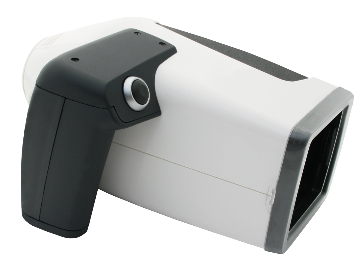
Figure 1: The Antera 3D® Handheld Optical Scanning Unit by Miravex Limited — Utilizing Photometric Stereo Technology for Quantitative Skin Analysis.
2. Feature Recommendation: The Antera 3D® Quantitative Ecosystem
For global procurement leaders seeking an industry-standard quantitative skin assessment device, the Antera 3D® system by Miravex Limited represents the benchmark in portable, high-precision dermatological metrology. Engineed in Dublin, Ireland, and backed by over 15 years of continuous optical hardware and algorithmic refinement, Antera 3D® delivers comprehensive multi-parameter quantitative profiling in under two seconds per acquisition.
Wrinkle & Texture Quantification
Measures surface roughness ($R_a$), root-mean-square roughness ($R_q$), maximum profile height ($R_z$), wrinkle depth, length, and overall skin indentation volumes with microscopic accuracy.
Melanin & Pigmentation Mapping
Isolates epidermal and dermal melanin distribution maps. Calculates hyperpigmentation cluster area, spot intensity concentration, and age-spot evolution across longitudinal studies.
Erythema & Vascular Analysis
Extracts hemoglobin absorption profiles to quantify micro-vascular redness, rosacea progression, post-inflammatory erythema (PIE), and inflammatory flare severity.
Pore & Depression Profiling
Automatically categorizes fine lines, open pores, and acne scars by depth ($mm$) and volume ($mm^3$), providing granular structural data for topical dermal treatments.
3D Topographical Surface Rendering
Renders true full-color 3D mathematical elevations, allowing research teams to view color-coded elevation maps, cross-sectional depth profiles, and lighting vector changes.
Automated Image Registration
Features spatial alignment software algorithms that automatically match baseline images with follow-up scans, eliminating operator positioning error across multi-month clinical trials.
2.1 Comparative Optical Engineering Matrix
To assist technical procurement officers and CRO clinical directors in performing vendor duediligence, the following comparative engineering matrix contrasts modern quantitative photometric stereo systems against traditional diagnostic apparatuses:
| Performance Metric | Photometric Stereo (Antera 3D®) | Traditional 2D Imaging Booths | Mechanical Contact Profilometers |
|---|---|---|---|
| Primary Measurements | Depth ($R_a, R_z$), Volume ($mm^3$), Melanin, Hemoglobin | 2D Pixel Area ($mm^2$), Uncalibrated RGB Color | Single Line Roughness Profile ($R_a$) |
| Spatial Resolution (X-Y) | Sub-micron ($< 0.1\,mm$) high-density sampling | Standard Camera Sensor Resolution | Discrete mechanical stylus spacing |
| Acquisition Speed | < 1.5 seconds per site scan | 3.0 to 10.0 seconds per capture | 2 to 5 minutes per linear scan |
| Operator Mobility | Portable Handheld Unit (< 1.0 kg) | Bulky Floor / Desk Station (> 25 kg) | Stationary Benchtop Laboratory Unit |
| Ambient Light Dependence | Zero (Sealed Controlled Contact Optics) | High (Requires dark room / hood encapsulation) | N/A (Contact probe) |
| Clinical Repeatability (CV) | Coefficient of Variation < 2.5% | Coefficient of Variation > 8.0% | Variable (High skin deformation risk) |