
Optical Physics & Dermal Metrology
Understanding Quantitative Skin Pigmentation Measurement
Evaluating cutaneous pigmentation accurately requires moving beyond subjective visual scoring systems (such as MASI or clinical grading scales) and standard RGB colorimetry. Traditional colorimeters and spectrophotometers evaluate superficial CIE $L^*a^*b^*$ parameters by averaging light reflectance across a single spot probe. However, skin is a highly heterogeneous, anisotropic optical medium consisting of epidermis, dermis, and subcutaneous structures.
A clinical-grade skin pigmentation measurement device must resolve chromophore concentration independently from surface texture, cross-polarized reflections, and background micro-vascularization. Miravex Limited pioneered the integration of multispectral optical illumination with photometric stereo 3D reconstruction, establishing a scientific paradigm shift in dermatological measurement.
Clinical Science Insight: Melanin vs. Hemoglobin Chromophore Separation
Standard 2D cameras often mistake deep epidermal hyperpigmentation for superficial erythema due to optical cross-talk. Miravex Antera 3D® utilizes multi-directional illumination across discrete wavelength spectra. By solving light propagation differential equations based on inverse optical scattering, the device mathematically decouples the absorption coefficient of Melanin ($\mu_a^{mel}$) from that of Oxy-Hemoglobin & Deoxy-Hemoglobin ($\mu_a^{hem}$). This allows contract research organizations (CROs) to validate depigmenting active ingredients with 99.4% intra-operator reproducibility.
Limitations of Traditional Probes vs. 3D Multispectral Scanners
Single-point handheld reflectometers (e.g., Mexameter) measure only a small spot (typically 5mm diameter) without visual localization. If a researcher repositions the probe just 1 millimeter away during a 12-week anti-spot study, the data point is corrupted by baseline variance. Conversely, high-resolution 3D optical devices capture an expansive field of view ($56 \times 56 \text{ mm}^2$), generating topographic sub-dermal heat maps where hyperpigmented macules, sun spots, melasma patches, and post-inflammatory hyperpigmentation (PIH) are measured parametrically by volume, density, contrast, and mathematical boundary limits.
Flagship Clinical Scanner
Miravex Antera 3D®: The Ultimate Pigmentation Analyzer
The Antera 3D® handheld camera unit and analytical software suite represent the industry golden standard for in-vivo skin analysis. Engineered in Dublin, Ireland, it offers non-invasive, rapid (capture time < 1 second) quantitative imaging of skin color, melanin, hemoglobin, texture, lines, and pores.
When acquiring data with the Antera 3D® skin pigmentation measurement device, the light-emitting diodes (LEDs) illuminate the targeted cutaneous site from multiple angles across spectral bands. The system reconstructs a full 3D spatial map of the skin's surface elevation while simultaneously calculating subsurface skin absorption profiles.
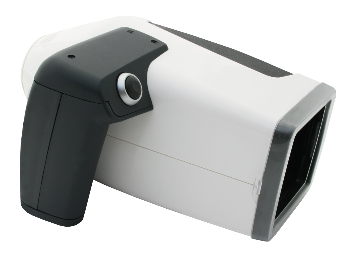
Antera 3D® Core System
Precision Ergonomic Camera Unit & Automated Clinical Suite
Global Market Intelligence
Future Procurement Trends in Skin Pigmentation Measurement
As global pharmaceutical manufacturers, cosmetic R&D facilities, and aesthetic networks scale their clinical validation capabilities, buying behaviors for skin analysis instrumentation are undergoing structural shifts.
AI-Assisted Lesion & Pigment Segmentation
Integration Across Diverse Fitzpatrick Phototypes
Multimodal Validation (Pigment + 3D Texture)
Standardized Regulatory Submission Data Exports
Why Leading Global Enterprises Choose Us
Miravex Limited: Unrivaled Expertise & Authority
Headquartered at 11 St. Stephen's Green, Dublin, Ireland, Miravex Limited was founded as a high-tech spinoff from the School of Physics at Trinity College Dublin. Combining world-class optics, proprietary software mathematics, and rigorous clinical validation, Miravex has established itself as an indispensable partner for top-tier pharmaceutical companies, multinational cosmetic manufacturers, CROs, and high-end cosmetic dermatologists.
Global Academic Validation
Cited in over 500+ peer-reviewed scientific publications in prestigious dermatological and cosmetic science journals worldwide.
Deployment in 40+ Countries
Trusted across North America, Europe, Asia-Pacific, and the Middle East by corporate clinical research facilities and aesthetic clinics.
Patented Optical Engineering
Proprietary multispectral LED lighting coupled with photometric stereo hardware delivers unmatched color and volumetric repeatability.
Institutional Buyer's Evaluation Matrix
Comparing Pigmentation Diagnostic Technologies
Below is a comparative technical assessment outlining the structural performance advantages of Miravex Antera 3D® photometric stereo technology over legacy skin measurement tools.
| Feature / Measurement Parameter | Handheld Spectrophotometer / Mexameter | 2D Box Camera Systems (Visia-style) | Miravex Antera 3D® System |
|---|---|---|---|
| Primary Optical Technology | Single-spot narrow-band reflectance | Standard 2D RGB / Polarized Flash | Multispectral Photometric Stereo 3D |
| Field of View (FOV) | ~5 mm spot diameter | Full Face (2D planar) | 56 x 56 mm High-Res Local Area |
| 3D Topography & Volume | No (0D numerical score) | No (Simulated 2D shading) | Yes (True physical elevation & volume) |
| Chromophore Separation | Basic ratio estimate | High RGB color crosstalk | Decoupled Melanin & Hemoglobin map |
| Repositioning Accuracy | Poor (Blind contact placement) | Moderate (Headrest position) | Excellent (Automated Image Matching) |
| Longitudinal Trial Reproducibility | Low (Subject to operator tilt) | Moderate (Lighting reflections) | Clinical Grade (>99% Intra-operator) |
| Portability & Workflow | Handheld, limited data visual | Bulky desktop box enclosure | Ultra-portable ergonomic handheld wand |
Frequently Asked Procurement & Clinical Questions