
1. Executive Summary & Paradigm Shift in Aesthetic Medical Diagnostics
The global medical aesthetics market is undergoing a fundamental structural transformation. Driven by heightened consumer scrutiny, stringent regulatory oversight, and AI-accelerated pharmaceutical formulation, the historical reliance on subjective visual scoring (such as the visual analog scale or qualitative photographic inspection) has become obsolete. Modern clinical practices and research institutions now demand empirical, highly reproducible biometric metrics. In this environment, the modern aesthetic medicine diagnostic device serves as the indispensable bridge between clinical intervention and quantitative validation.
Leading medical aesthetic diagnostic platforms have evolved beyond conventional 2D planar photography. Contemporary diagnostic devices incorporate optical multispectral illumination, photometric stereo reconstruction, and mathematical subsurface signal processing. By generating high-resolution 3D dermal topographies and isolating optical absorption spectra, these instruments quantify epidermal texture, volumetric alterations, melanin distribution, and hemoglobin concentration with micrometer precision.
For procurement officers, medical directors, and principal investigators, choosing the correct diagnostic platform directly impacts multi-center clinical trial validity, patient retention rates, treatment conversion ratios, and long-term brand equity. This document outlines the optical physics, clinical applications, procurement criteria, and future technological trajectories governing the aesthetic medicine diagnostic device market.
"The integration of objective, 3D multispectral imaging in aesthetic diagnostic devices has transformed clinical trial endpoints from qualitative estimations into repeatable mathematical constants." — Dr. Charles Taylor, Optical Physics Consultant.
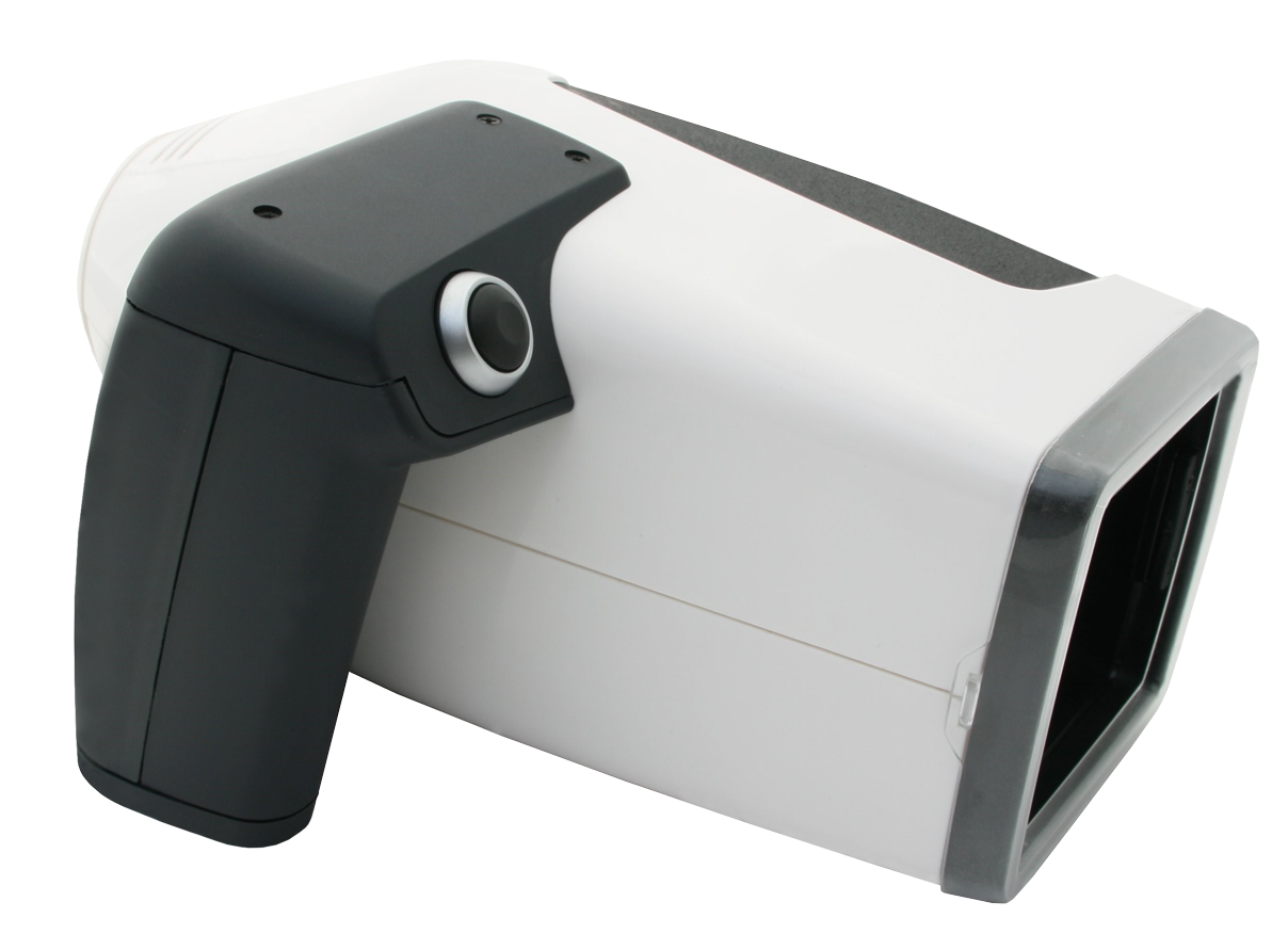
Figure 1: The Antera 3D® handheld multispectral aesthetic medicine diagnostic device engineered by Miravex Limited.
2. Core Technical Architecture of Modern Aesthetic Medicine Diagnostic Devices
To evaluate the diagnostic accuracy of modern dermal scanners, enterprise procurement teams must understand the underlying optical hardware and computational software architectures. Standard commercial facial scanners often fail under clinical scrutiny due to uncontrolled ambient lighting, positioning variations, and surface light reflection artifacts. Purpose-built aesthetic medicine diagnostic devices solve these physics challenges through three core engineering pillars:
2.1 Photometric Stereo Reconstruction
Photometric stereo technology utilizes multiple illumination sources directed at pre-calibrated, distinct incident angles toward the targeted tissue surface. By acquiring multiple sequential images under varying lighting angles while maintaining a fixed camera perspective, proprietary algorithms solve the reflectance map equations (reflectance map gradient space). This allows the diagnostic device to mathematically calculate the surface normals at every pixel, reconstructing true 3D micro-topography with sub-micron elevation resolution. Unlike fringe projection systems or laser scanners, photometric stereo allows handheld portability while capturing sub-millimeter skin topography without mechanical drift.
2.2 Multispectral LED Array & Polarized Light Filtering
Biological skin tissue exhibits wavelength-dependent optical absorption and scattering properties. Advanced diagnostic devices feature custom LED illumination chips spanning visible light spectrums (from 365 nm UV-A to near-infrared 850 nm). Furthermore, cross-polarization filters decouple specular surface reflection (glare from stratum corneum lipids and moisture) from diffuse subsurface reflectance. This enables clean optical penetration into the upper dermis, isolating light absorbed specifically by target chromophores: melanin in the basal layer and hemoglobin in the papillary dermal microvascular network.
2.3 Quantitative Chromophore Mapping (Beer-Lambert Law Derivative Algorithms)
By processing multispectral reflectance data through modified Beer-Lambert equations, the device separates pigmentation signals from vascular redness. Rather than offering arbitrary color-matching RGB values, enterprise diagnostic devices generate quantitative concentration maps representing true melanin density (expressed in relative concentration indices) and hemoglobin concentration (erythema intensity and capillary dilation).
| Diagnostic Metric | Conventional 2D Systems | Advanced 3D Multispectral Devices (e.g. Antera 3D®) |
|---|---|---|
| Wrinkle & Depression Analysis | Subjective 2D shadow visualization; susceptible to camera angle bias | 3D depth, volume (mm³), average roughness (Ra, Sa), maximum indentation depth |
| Pigmentation Assessment | RGB color brightness; cannot distinguish superficial from deep melanin | Multispectral melanin concentration maps; true optical absorption indexing |
| Redness & Vascularity | Red pixel channel extraction; distorted by skin tone and ambient flash | Subsurface cross-polarized hemoglobin concentration mapping; erythema quantification |
| Volume Changes (Fillers/Scars) | Impossible; relies on visual estimation | 3D volumetric differential matching (mm³ matrix comparison pre- and post-treatment) |
| Ambient Light Dependence | High distortion; requires dedicated darkroom conditions | Self-shielded optical dome with closed-loop illumination calibration |
3. Recommended Platform: Antera 3D® Diagnostic Device Architecture
When benchmarking market solutions against rigorous E-E-A-T (Experience, Expertise, Authoritativeness, and Trustworthiness) criteria, the Antera 3D® developed by Miravex Limited stands out as an industry reference standard across global cosmetic research laboratories and clinical aesthetic practices.
3.1 Modular Application Capabilities
Engineered for multi-disciplinary versatility, the platform supports dedicated software modules tailored to specific research and clinical endpoints:
- Wrinkles & Texture Analysis Module: Calculates fine-line density, skin roughness parameters ($Ra$, $Rq$, $Sa$, $Sq$), depression volume ($mm^3$), and contour orientation. Crucial for assessing anti-aging topical efficacy, injectable neuromodulators (Botulinum Toxin), and fractional laser resurfacing outcomes.
- Pigmentation & Sun Damage Module: Maps hyperpigmentation, melasma, solar lentigines, and post-inflammatory hyperpigmentation (PIH). Measures average melanin concentration and spatial uniformity coefficient, supporting depigmenting active ingredient validation and IPL therapy tracking.
- Redness & Vascularity Module: Isolates rosacea, telangiectasia, erythema, and inflammatory lesions. Measures hemoglobin concentration, lesion surface area, and vascular density before and after vascular laser (V-Beam, KTP) or anti-inflammatory treatments.
- Pores & Micro-Relief Mapping: Measures pore volume, diameter, depth, and spatial distribution count. Essential for sebum-control topicals, microneedling therapies, and chemical peeling evaluation.
- 3D Volumetric Matching Module: Offers automated alignment of baseline and follow-up scans. Quantifies structural volume gain or loss in cubic millimeters ($mm^3$), providing verified visual proof for dermal filler augmentation, scar revision, and lipo-filling procedures.
Explore Precision Dermal Diagnostics
Request technical specifications, clinical validation studies, and direct enterprise pricing for the Antera 3D® diagnostic device.
Send an Inquiry4. Future Procurement & Technological Trends in Aesthetic Medicine Diagnostic Devices (2025–2030)
Global procurement managers and clinical trial coordinators must anticipate upcoming technological shifts when investing in diagnostic infrastructure. Over the next five to ten years, several key macro trends will redefine the aesthetic medical diagnostic device landscape:
4.1 AI-Driven Biometric Predictive Aging & Generative Simulation
Artificial intelligence is moving beyond rudimentary image recognition into generative predictive biometrics. Next-generation diagnostic devices will combine historical 3D topography databases with machine learning models to simulate five-to-ten-year patient aging trajectories under varying environmental exposure scenarios. Furthermore, AI algorithms will generate realistic post-treatment visual forecasts based on multi-modal clinical input (e.g., combining filler rheology data with skin elasticity metrics).
4.2 Cloud-Integrated Multi-Center Clinical Trial Standardization
Pharmaceutical companies, contract research organizations (CROs), and multinational cosmetic brands are increasingly centralizing their R&D data. Diagnostic devices are shifting toward cloud-native ecosystems with automated cryptographic hashing for regulatory audit trails (FDA 21 CFR Part 11 compliance). Multi-site trial managers can instantly harmonize optical measurement metrics captured across clinical sites in North America, Europe, and Asia-Pacific, eliminating inter-operator variability.
4.3 Non-Invasive Sub-Dermal Structural Biomarker Profiling
Future diagnostic platforms will integrate sub-surface optical coherence tomography (OCT) or multiphoton imaging concepts into compact handheld form factors. This evolution will enable non-invasive quantification of dermal collagen density, elastin fiber orientation, and epidermal basement membrane thickness in real-time, removing the requirement for invasive punch biopsies in cosmetic efficacy research.
4.4 ROI-Optimized Consultation Workflows in Aesthetic Clinics
In commercial aesthetic clinics, diagnostic devices are becoming central sales enablement instruments. Patients demand transparency; interactive 3D visualizations showing quantitative percentage improvements (e.g., "24.6% reduction in deep wrinkle volume after 3 treatment sessions") directly elevate treatment plan acceptance rates, patient satisfaction scores, and lifetime customer value (LTV).
5. Enterprise Advantage & Proven Authority (E-E-A-T Excellence)
Selecting an aesthetic medicine diagnostic device supplier requires rigorous evaluation of enterprise credibility and technological heritage. Miravex Limited, headquarterd in Dublin, Ireland, embodies the highest standards of scientific rigor and market validation.
5.1 Deep Scientific & Academic Heritage
Spun out of the renowned School of Physics at Trinity College Dublin, Miravex was founded on decades of advanced optical research in light transport within complex scattering media. The company’s patented technologies represent direct translations of high-energy optical physics into practical clinical dermatological applications.
5.2 Over 500 Peer-Reviewed Scientific Publications
The Antera 3D® system is not merely a commercial marketing tool; it is a validated scientific instrument. It has been cited in over 500 peer-reviewed medical and dermatological journal articles globally—including the Journal of Cosmetic Dermatology, Lasers in Surgery and Medicine, Dermatologic Surgery, and Archives of Dermatological Research. This extensive body of independent validation guarantees that the data produced meets international clinical trial standards.
5.3 Global Deployment in 40+ Countries
Deployed in more than 40 countries worldwide, Miravex systems are trusted by top-tier global pharmaceutical conglomerates (such as Galderma, L'Oréal, Procter & Gamble, Unilever, Johnson & Johnson, and Merz Aesthetics), leading university hospitals, and prestigious private aesthetic medical clinics.
5.4 Medical-Grade Manufacturing & Calibration Reliability
Manufactured under ISO 13485 quality management standards, each Antera 3D® unit undergoes rigorous factory spectral and spatial calibration. The system’s enclosed optical design ensures immunity to ambient light fluctuations, guaranteeing intra-operator reproducibility and long-term instrument stability across multi-center clinical trials.
6. Global B2B Buyer Frequently Asked Questions (FAQ)
Below are answers to critical technical and procurement questions commonly raised by corporate buyers, research directors, and clinical administrators when evaluating aesthetic medicine diagnostic devices.
Q1: What makes a 3D multispectral aesthetic medicine diagnostic device superior to standard 2D facial photography units?
Standard 2D photography captures flat RGB light reflections, which are heavily distorted by ambient room lighting, flash intensity, patient head rotation, and facial expressions. 2D images cannot measure spatial depth or volume. In contrast, a 3D multispectral diagnostic device (such as Antera 3D®) uses controlled directional illumination and cross-polarized light to isolate target skin layer chromophores (melanin and hemoglobin) and construct quantitative, micrometer-accurate 3D topographies that are unaffected by external room lighting.
Q2: How does an aesthetic diagnostic device quantify dermal hemoglobin and melanin concentrations independently?
The device emits specific narrow-band wavelengths of light using a high-density LED matrix. Because melanin and hemoglobin possess distinct light absorption spectra, cross-polarized optical sensors capture the reflected light and apply mathematical algorithms derived from the Beer-Lambert law. This mathematical decoupling allows the system to extract precise, separate concentration maps for melanin (pigmentation) and hemoglobin (vascular redness) without cross-channel signal bleeding.
Q3: Is the Antera 3D® diagnostic device suitable for both clinical aesthetic consultation and CRO pharmaceutical research?
Yes. Miravex offers software suites tailored to both operational environments. For clinical practices, Antera 3D® CS provides rapid patient scanning, intuitive visual before-and-after consultation screens, and quick report generation to boost treatment compliance. For CROs, biotech institutions, and cosmetic R&D laboratories, Antera 3D® Lab provides raw data exporting ($Ra$, $Sa$, volume in $mm^3$, CIELAB color space values), batch processing capabilities, statistical analysis tools, and multi-site database synchronization.
Q4: What specific optical metrics are measured during wrinkle and skin texture analysis?
The diagnostic system measures mathematical surface roughness parameters in accordance with international ISO surface metrology standards. Key metrics include average roughness ($Ra$), root-mean-square roughness ($Rq$), 3D area roughness ($Sa$, $Sq$), maximum indentation depth ($mm$), surface area index, and total depression volume ($mm^3$). These objective biometrics enable quantitative tracking of skin smoothing over precise time intervals.
Q5: How does ambient lighting affect optical measurement accuracy during clinical operation?
With the Antera 3D® device, ambient light interference is completely eliminated. The unit features an ergonomically designed contoured soft aperture dome that seals gently against the subject’s skin surface during capture. This creates a fully enclosed, light-tight optical cavity. Combined with closed-loop LED flash control, measurements are 100% reproducible regardless of ambient room lighting conditions or geographic clinical site variations.
Q6: Can the diagnostic device track volume changes resulting from dermal fillers, fat grafting, or scar reconstruction?
Yes. Using advanced 3D surface matching algorithms, the software aligns baseline scans with post-treatment scans using fixed anatomical landmarks. It calculates localized volume changes in cubic millimeters ($mm^3$), displaying dynamic color-coded elevation maps (showing green/blue for volume addition and red/yellow for volume reduction). This capability is critical for validating injectable volumizers, lipo-transfer stability, and hypertrophic/atrophic scar therapies.
Q7: What are the regulatory standards and data export options for international cosmetic clinical trials?
Antera 3D® software supports multi-format data export options (including CSV, Excel, high-resolution TIFF, JPEG, and 3D OBJ mesh formats) for seamless integration with statistical analysis software (such as SPSS, SAS, or R). The software architecture complies with international clinical data integrity guidelines, enabling cryptographic logging required for multi-center global clinical trials.
Q8: What is the typical ROI timeline for installing an aesthetic medicine diagnostic device in a commercial med-spa or clinic?
Most commercial aesthetic practices achieve full capital equipment payback within 3 to 6 months. By presenting objective, quantitative proof of skin damage during initial consultations, clinics experience an immediate increase in treatment plan conversion rates (often between 25% to 40%). Furthermore, objective follow-up scans document subtle micro-improvements that patients might otherwise miss, dramatically improving patient retention, referral rates, and repeat procedure bookings.
Q9: How frequently does the optical hardware require re-calibration or factory maintenance?
The Antera 3D® is built with solid-state LED illumination and durable optical components designed for high-throughput daily use. The platform includes a quick, user-executable optical calibration target plate and software routine that takes less than one minute to perform. Routine user calibration ensures instrument precision over extended operational lifespans without requiring frequent factory recalibration.
Q10: How does the software integrate with existing clinic management software or Electronic Medical Record (EMR) systems?
The Antera 3D® software environment allows centralized patient database storage on local clinic networks or secure enterprise cloud servers. Patient records, diagnostic images, and generated PDF reports can be exported directly or linked into standard practice management platforms and electronic medical record systems via standardized file structures.
7. Conclusion & Global Procurement Consultation
Investing in a premium aesthetic medicine diagnostic device is a strategic decision that elevates clinical credibility, scientific accuracy, and commercial ROI. Whether you are validating next-generation active pharmaceutical ingredients, conducting rigorous CRO clinical trials, or growing a top-tier aesthetic dermatology practice, the Antera 3D® platform delivers the quantitative precision, E-E-A-T background, and technological reliability modern medicine requires.
Schedule a Technical Demonstration or Request a Quote
Speak directly with our clinical application specialists and global distribution team to discuss your practice or research requirements.
Send an Inquiry