
Industrial Skin Texture Assessment & 3D Imaging Systems
Our turnkey OEM/ODM hardware and software platforms offer quantitative optical diagnostic capabilities engineered to scale from portable diagnostic tools to enterprise clinical scanners.


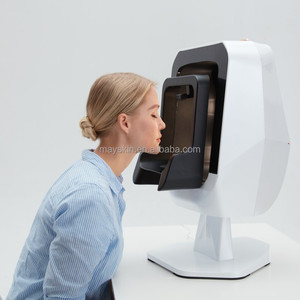
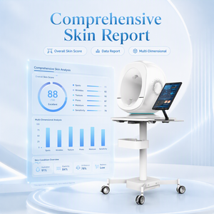
The Physics of Quantitative Skin Texture Assessment: Photometric Stereo & Micro-Topography
Modern clinical dermatology and cosmetic claim validation have shifted permanently from subjective visual scoring to objective, metrology-grade volumetric metrics. As a premier OEM/ODM Skin Texture Assessment Machine Manufacturer & Exporter, our engineering framework integrates advanced Photometric Stereo Imaging with high-resolution directional lighting arrays to measure skin surface micro-relief with micron-level reproducibility.
Conventional two-dimensional digital photography is fundamentally limited by environmental illumination drift, specular highlights, and a lack of depth perception. Our proprietary photometric engine captures a sequence of images under directional illumination from multiple calibrated LED angles (ranging from cross-polarized parallel lights to narrow-band UV and deep-blue illuminants). By analyzing the intensity variations across varying directional illumination vectors, the system reconstructs the local surface normal vectors for every single pixel across the target anatomical zone.
Technical Information Gain: By mathematically integrating local surface normals, our 3D processing algorithm constructs a true spatial micro-topography mesh independent of skin pigmentation or ambient lighting artifacts. This allows researchers to isolate real physical structural variations—such as fine line volumetric depth, pore rim height, and hyper-keratosis roughness—from optical color variations.
ISO 25178 Standardized Surface Roughness Metrics
To provide contract research organizations (CROs) and pharmaceutical laboratories with publication-ready dataset compliance, our internal software pipeline translates 3D mesh points into standardized ISO 25178 areal roughness parameters:
- Sa (Areal Average Roughness): Measures the absolute height difference across the micro-relief topography, establishing baseline smoothness metrics for anti-aging formulation trials.
- Sq (Root Mean Square Height): Quantifies structural variances, providing extreme sensitivity when tracking subtle micro-wrinkle attenuation over 14-day to 90-day clinical intervention periods.
- Sz (Maximum Surface Height): Tracks deep furrow depth, scarring structural features, and deep expression lines with sub-micron vertical resolution.
- Sp & Sv (Peak Height & Pit Depth): Separates surface papule formations (e.g., acne lesions or follicular congestion) from localized atrophy or pore enlarged indentations.
By offering full SDK control over illuminant pulse sequences, exposure control, and raw uncompressed sensor output (14-bit RAW format), our OEM hardware platform enables medical device brands and biotech entities to embed custom quantitative algorithms directly into their proprietary diagnostic software ecosystem.
Enterprise OEM/ODM Engineering & Customization Capabilities
Building a commercial-grade medical or cosmetic skin analyzer requires striking a precise balance between optical accuracy, mechanical ergonomics, thermal stability, and regulatory compliance. As a vertically integrated exporter and original design manufacturer, we maintain comprehensive in-house R&D divisions spanning optical engineering, industrial design, firmware architecture, and AI algorithm development.
Subsurface Chromophore Mapping: Deconvolution of Hemoglobin and Melanin
Skin texture cannot be analyzed in isolation from the underlying dermal matrix. Structural breakdown—such as collagen degradation and elastin loss—is frequently accompanied by vascular dysregulation (erythema, telangiectasia) and irregular melanogenesis (hyperpigmentation, solar lentigines).
Our OEM skin texture machines integrate dual-band cross-polarized spectral deconvolution. By isolating specific absorption spectrums of epidermal melanin ($400-500\text{ nm}$) and dermal hemoglobin ($540-580\text{ nm}$), our machines produce separate quantitative heat maps of:
- Subsurface Erythema Distribution: Mapping localized inflammation, post-inflammatory hyperpigmentation (PIH) vulnerability, and rosacea severity index.
- Melanin Density Mapping: Differentiating superficial epidermal pigmentation from deep dermal melasma, allowing clinicians to select precise laser wavelengths or chemical peeling protocols.
- Pore Congestion & P. Acnes Biofilms: Utilizing $405\text{ nm}$ narrow-band UV fluorescence to excite bacterial porphyrins, revealing active acne flare-up zones prior to visual clinical outbreak.
Comparative Analysis: Advanced Photometric 3D vs. Standard RGB Systems
When selecting an OEM manufacturing partner, procurement directors and product managers must evaluate the optical limitations of standard commercial scanners against medical-grade micro-topography platforms:
| Evaluation Parameter | Standard Commercial RGB Scanners | Industrial OEM 3D Photometric Platform |
|---|---|---|
| Primary 3D Method | Software-interpolated simulated shadows (Low accuracy) | Photometric Stereo Vector Mesh Integration |
| Vertical Resolution ($Z$-Axis) | Uncalibrated / Arbitrary Pixel Units | Calibrated to ±2.5 µm Absolute Metric Scale |
| Roughness Standard | Visual relative scoring (1–10 scale) | ISO 25178 Quantitative Metrics (Sa, Sq, Sz) |
| Illumination Rig | Single ring light or unpolarized ambient LED | Multi-angle cross-polarized narrowband LED array |
| Subsurface Chromophores | Color channel separation (RGB color space) | Spectral Deconvolution (Melanin / Hemoglobin) |
| SDK & Enterprise White-Label | Locked consumer software app | Full Modular C++/Python SDK & Raw Hardware Access |
Procurement Trends & Technological Roadmap (2026–2030)
As the global medical aesthetics and dermatological research markets expand toward an estimated $28.5 Billion valuation by 2030, procurement requirements for hardware distributors, pharmaceutical brands, and clinical trial networks are undergoing a fundamental transformation. B2B buyers must align their product development pipelines with three macro trends driving the next decade of diagnostic hardware:
1. Transition from Qualitative Imaging to AI-Driven Longitudinal Tracking
Historical skin analysis relied heavily on static "before-and-after" photographs taken weeks apart. Current enterprise buyers demand dynamic longitudinal alignment algorithms. Modern systems utilize 3D facial feature point landmarking (extracting over 120 facial coordinates) to automatically register, scale, and map identical skin patches ($10\text{mm} \times 10\text{mm}$) across multiple visits. This guarantees that changes in micro-texture ($R_a$) reflect true therapeutic efficacy rather than minor positioning variations.
2. Modular Platform Architectures and Cloud-Native SDK Integration
Hardware isolation is no longer acceptable for chain aesthetic clinics or international CRO trials. Contemporary procurement specifications require hardware to function as a edge node within broader cloud telemetry ecosystems. Our OEM platforms feature embedded processing units capable of executing real-time image normalization and local tensor calculations, transmitting lightweight structured JSON payloads and compressed 3D OBJ mesh files directly to centralized HIPAA/GDPR-compliant cloud data lakes.
3. Multi-Modal Optical Diagnostic Fusion
The future of skin texture evaluation lies in combining surface micro-topography with functional optical measurements. Next-generation ODM pipelines are integrating high-frequency optical coherence tomography (OCT) and hyperspectral sensors within handheld form factors. This multi-modal approach enables simultaneous evaluation of stratum corneum hydration levels, dermal collagen fiber density, and epidermal surface roughness in a single non-invasive scan.