
Featured Dermatoscope Imaging Systems
Engineered to international CE standards, incorporating cross-polarized filters, multi-spectral LED emission arrays, and clinical-grade 3D surface topography software.
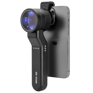
- Dual-mode Cross & Parallel Polarization
- 365nm Narrowband UV Pigment Illumination
- 10X Precision Optical Achromatic Lens
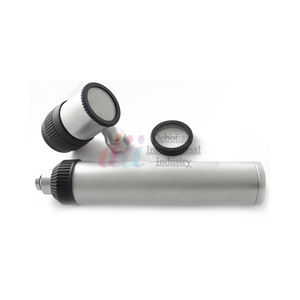
- Surgical Stainless Steel / Manual Metal Casing
- Uniform Fiber-Optic Light Transmission
- ENT & Dermatology Multi-speculum Adaptability
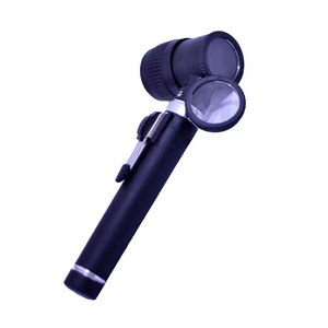
- Achromatic Triplet Glass Element Array
- Scale-Calibrated Contact Plate Grid
- Heavy-Duty Aluminum Protective Suite
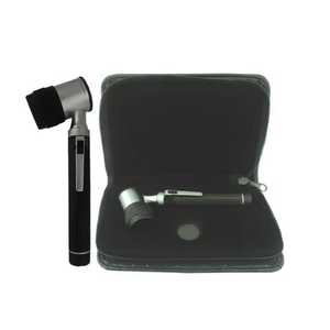
- Constant-Current LED Driver Circuitry
- High-Impact Ergonomic Polymer-Steel Housing
- Distortion-Free Edge-to-Edge Field of View
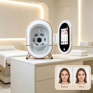
- Photometric Stereo 3D Surface Reconstruction
- WiFi Cloud PACS Data Synchronization
- AI Lesion Pattern Recognition Engine
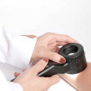
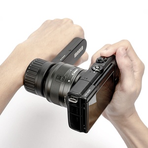
- Universal Smartphone Magnetic Mount Compatibility
- Zero-Spherical Aberration Multi-Element Lens
- Lithium-Ion Fast Charge Power Architecture
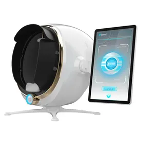
- Full Facial Topography & Pore Mapping
- Subsurface Vascular Erythema Quantification
- Quantitative Wrinkle Volumetric Depth Metrics
- Aircraft-Grade Anodized Aluminum Alloy Body
- Stepless Light Intensity Brightness Dimmer
- Autoclavable Glass Contact Faceplate
The Evolution of Optical Dermoscopy Optics
In modern dermatology, clinical efficacy, diagnostic sensitivity, and skin cancer screening precision are fundamentally governed by optical engineering standards. The transition from basic glass magnifiers to advanced multi-spectral cross-polarized dermatoscopes has redefined clinical diagnostic accuracy for melanocytic lesions, basal cell carcinomas (BCC), dysplastic nevi, and vascular anomalies.
When light strikes human skin, approximately 4% to 7% of incident light is immediately reflected back by the stratum corneum due to the refractive index step between air ($n \approx 1.0$) and keratin ($n \approx 1.55$). This specular glare obscures deeper architectural features within the epidermis, dermo-epidermal junction (DEJ), and upper papillary dermis. Polarized dermatoscope optics isolate deep-diffuse backscattered light, enabling visualization of vascular networks, pigment networks, chrysalis structures, and regression zones.
Cross-Polarization Physics
Utilizes dual linear polarizers set perpendicular to each other. Reflected specular light maintains polarization state and is blocked by the analyzer, while photons penetrating deep into the dermis depolarize and reach the optical sensor.
Multi-Spectral 365nm UV
Emits narrow-spectrum ultraviolet illumination (365nm) to induce epidermal and dermal fluorophore excitation. Crucial for assessing superficial hyperpigmentation, porphyria, acne propionibacterium, and localized collagen breakdown.
Photometric 3D Topography
Combines multi-angle directional LED lighting arrays with mathematical photometric stereo algorithms to compute sub-micron 3D surface topography, measuring wrinkle volume, pore dimensions, and skin roughness quantitatively.
Optical Dermatoscope Future Development Trends
1. Edge AI Deep Learning Integration
Next-generation dermatoscope imaging hardware directly embeds neural network microprocessors. Real-time automatic feature extraction highlights asymmetry, border irregularity, color variation, and differential structural diameter (ABCD rule metrics) directly on the viewer screen.
2. Mobile Tele-Dermoscopy Ecosystems
B2B buyers are demanding modular smartphone clip-on architectures equipped with universal magnetic alignment plates. High-grade optical glass lenses allow primary care practitioners to capture studio-grade clinical images and upload them seamlessly into hospital cloud PACS system.
3. Hyperspectral & Biomarker Mapping
The shift from broad RGB spectrum illumination to narrow-band multi-spectral light sources (ranging from 365nm UV to 940nm Near-Infrared) allows clinicians to isolate subsurface hemoglobin concentration maps and melanin spatial distribution without invasive skin biopsies.
4. Quantitative 3D Surface Topography
Integrating 3D photometric stereo imaging into desktop dermatoscope analyzers allows cosmetic manufacturers, CROs, and aesthetic surgeons to measure wrinkle depth in microns, pore volumetric changes, and skin smoothness post-laser treatment with clinical statistical validity.
Technical Specification Benchmark Matrix
| Optical Parameter | Standard Handheld Dermatoscope | Pro Cross-Polarized Model (e.g. DE-4100) | Clinical 3D Desktop Scope System |
|---|---|---|---|
| Optical Magnification | 10X Fixed Zoom | 10X Achromatic Triplet System | 10X to 50X Dynamic Digital/Optical Zoom |
| Illumination Spectrum | White LED (450nm-650nm) | Dual-Mode White LED + 365nm Narrowband UV | Multi-Spectral Directional LED Array |
| Polarization Efficiency | Single Linear Filter (>85%) | Cross-Polarized Field (>99.4%) | 3D Photometric & Polarized Hybrid Grid |
| Lens Coating & Glass | Standard Optical Glass | Schott Glass with Broadband Anti-Reflective Coating | Multi-Element Low Dispersion Optical Lens System |
| Sensor & Output | Optical Eyepiece Only | Smartphone Universal Magnetic Interface | High-Res CMOS Sensor + WiFi Cloud PACS Interface |
| Regulatory Standards | CE, RoHS | CE Standard, ISO 13485, IEC 60601-1-2 EMC | CE Medical, FDA Class II, ISO 13485 Certified |
Why Partner with Our Optical Manufacturing Hub
ISO 13485 & Cleanroom Production
Our manufacturing facility operates state-of-the-art Class 10,000 dust-free optical assembly cleanrooms. Every optical lens pair undergoes laser alignment checks to guarantee zero spherical and chromatic aberration across all zoom spectrums.
Complete OEM/ODM Customization Matrix
We support international B2B distributors with comprehensive OEM/ODM services: private label laser engraving, bespoke optical filter bandwidth customization, custom software algorithms, and tailored packaging configurations.
Global Supply Chain & Export Reach
Exporting to over 40 countries, our logistics network provides seamless door-to-door B2B deliveries, comprehensive customs compliance documentation, CE certification, and rapid turnaround for volume production orders.