
Flagship Subdermal Vascular & Dermal Analysis Systems
Explore China's top-tier OEM/ODM spectral facial imaging systems engineered for aesthetic clinics, CROs, research institutes, and medical spas.


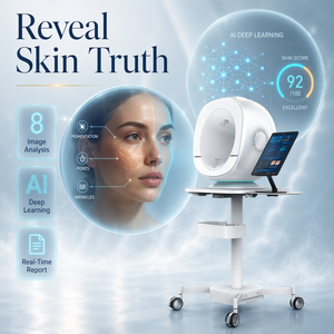



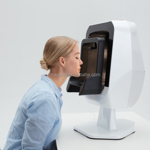
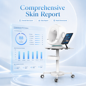
Executive Industry Report: Subdermal Vascular Imaging Manufacturing Landscape in China
In contemporary aesthetic medicine, clinical dermatology, and pharmaceutical cosmetics research, non-invasive quantitative measurement of dermal architecture has undergone a paradigm shift. Subdermal vascular imaging devices—utilizing multi-spectral cross-polarized illumination, photometric stereo 3D reconstruction, and AI-driven optical coherence algorithms—represent the bleeding edge of sub-surface tissue analysis. As medical aesthetic institutions move away from subjective visual assessments, the global demand for high-precision dermal imaging hardware manufactured by specialized Chinese factories has experienced exponential growth.
China has emerged as the global epicenter for precision optical manufacturing, medical-grade CMOS sensor integration, and artificial intelligence diagnostic software development. Leading Chinese manufacturers now supply over 65% of the world’s sub-surface vascular analysis hardware under both proprietary brands and specialized OEM/ODM agreements. This whitepaper offers an exhaustive technical evaluation of China's top subdermal vascular imaging device factories, analyzing optical physics, manufacturing quality frameworks, software SDK capabilities, and strategic procurement methodologies for international buyers, distributors, and contract research organizations (CROs).
Core Industry Insight: Next-generation subdermal imaging transcends superficial RGB skin photography. By isolating specific absorption peaks of hemoglobin (Hb) and oxyhemoglobin (HbO2) at 540nm and 577nm wavelengths, modern Chinese diagnostic hardware maps vascular erythema, telangiectasia, and microcirculatory inflammation deep within the papillary and reticular dermis with micrometer-scale resolution.
Technical Capabilities of Leading Chinese Manufacturing Hubs
The industrial cluster for advanced biophotonics in China—primarily concentrated in Shenzhen, Suzhou, Wuhan, and Shanghai—leverages robust vertical integration. Factories in these regions maintain full-stack control over optical lens polishing, LED spectral array design, PCB assembly, and cloud-based AI algorithmic modeling. This integrated ecosystem ensures that international procurement partners receive devices complying with rigorous international standards, including ISO 13485, CE Medical Devices Directive (MDD/MDR), US FDA Class II guidelines, and RoHS environmental compliance.
Subdermal Vascular Optical Physics: How Multispectral Analysis Works
Understanding the optical physics underlying modern subdermal vascular imaging is essential for procurement managers evaluating factory claims. Human skin consists of three primary optical layers: the stratum corneum/epidermis, the dermis, and subcutaneous tissue. Light striking the skin undergoes reflection, refraction, scatter, and selective absorption by chromophores—predominantly melanin in the epidermis and hemoglobin within the vascular network of the dermis.
Standard RGB imaging cameras capture visible light reflected primarily from the skin surface (epidermis). In contrast, specialized subdermal vascular scanners manufactured in China utilize narrowband spectral illumination combined with cross-polarization techniques.
1. Cross-Polarized Illumination Architecture
When linearly polarized light strikes the skin surface, approximately 4% to 7% is reflected directly back with its polarization state intact (specular glare). The remaining light penetrates into the dermal layers, undergoing multiple scattering events that randomize its polarization state before backscattering out of the skin (diffuse reflectance).
By placing a linear polarizer in front of the light source and a second polarizer (analyzer) rotated 90 degrees in front of the CMOS camera sensor, Chinese imaging devices block specular glare completely. This allows the system to capture only the diffusely scattered light that has interacted directly with subsurface microvasculature, providing crisp, glare-free visualization of capillaries, spider veins, and deep inflammation patterns.
2. Spectral Chromophore Decomposition (Hemoglobin vs. Melanin)
Different biological chromophores display unique optical absorption spectra. Oxyhemoglobin (HbO2) exhibits sharp absorption peaks around 415nm (Soret band), 542nm, and 577nm (Q-bands). Deoxyhemoglobin (Hb) exhibits a single peak near 555nm. Melanin, on the other hand, demonstrates a monotonic decrease in absorption as wavelength increases from UV (365nm) into the near-infrared (NIR) spectrum (850nm+).
| Wavelength / Spectrum | Target Dermal Depth | Primary Diagnostic Target | Clinical & Aesthetic Application |
|---|---|---|---|
| 365nm (Standard UV) | Epidermal Layer (< 0.1mm) | Porphyrins, Sebum, UV Spots | Acne vulgaris mapping, photodamage evaluation |
| 415nm (Deep Violet / Blue) | Superficial Papillary Dermis (0.1 - 0.2mm) | Superficial Capillary Loops, Propionibacterium | Rosacea diagnosis, early inflammation assessment |
| 542nm / 577nm (Green Band) | Mid Dermis (0.3 - 0.6mm) | Oxyhemoglobin (HbO2) Vessels | Telangiectasia, erythema quantification post-laser |
| 630nm / 660nm (Red Band) | Deep Dermis (0.8 - 1.2mm) | Deep Vascular Plexus, Structural Collagen | Vascular lesion mapping, deep scar tissue analysis |
| 850nm (Near-Infrared / NIR) | Subcutaneous Layer (> 1.5mm) | Feeder Veins, Venous Malformations | Sclerotherapy navigation, plastic surgery vascular mapping |
Advanced Chinese factory models—such as the X5PRO 9-Spectral and A6 3D Facial Scanner featured above—incorporate mathematical matrix decomposition algorithms. By analyzing pixel-by-pixel attenuation across multiple narrow wavelength bands, the internal DSP (Digital Signal Processor) isolates the distinct absorption profiles of melanin and hemoglobin. This generates pure, unmixed red-map (vascular) and brown-map (pigment) diagnostic images with absolute quantitative confidence.
Global Sourcing & Technology Evolution Trends (2026–2030)
As international distributors, aesthetic chain clinics, and medical equipment brands establish supply chain partnerships in China, understanding future technology trajectories is paramount. Over the next five years, the subdermal imaging landscape will evolve rapidly across three primary technological vectors:
1. Hyper-Fast Multi-Spectral Frame Acquisition
Legacy skin analyzers required 10 to 15 seconds to cycle through mechanical filter wheels or sequentially strobe LED arrays. Modern OEM/ODM factories in China are transitioning to high-speed solid-state LED arrays synchronized with global-shutter CMOS sensors. Modern devices capture up to 9 distinct spectral channels in less than 0.8 seconds. This rapid capture minimizes micro-motion artifacts caused by patient breathing or subtle head tremors, yielding ultra-sharp, sub-pixel accurate vascular alignment.
2. Integration of Embedded Generative AI & Predictive Modeling
Hardware alone is no longer the sole differentiator. Leading Chinese exporters are now embedding local NPU (Neural Processing Unit) hardware directly into device mainboards. This enables local execution of generative aging algorithms, treatment simulation models (e.g., predicting vascular clearance post-IPL or Nd:YAG laser therapy), and automated clinical progress monitoring without sending patient data to third-party cloud servers—a critical feature for GDPR and HIPAA compliance.
3. Miniaturization & Handheld Point-of-Care Systems
While benchtop facial scanners remain the gold standard for full-face consultations, there is a surging procurement demand for portable, handheld multi-spectral dermoscopes. Chinese exporters are pioneering ultra-compact optical modules that clip onto tablets or operate wirelessly via high-speed Wi-Fi 6 protocols, bringing clinical-grade vascular analysis to bedside dermatology, mobile cosmetic consultations, and boutique clinical trials.
China OEM/ODM Factory Infrastructure & Quality Control Standards
Evaluating a manufacturing partner requires looking beyond glossy product catalogs. Top China subdermal vascular imaging device exporters possess sophisticated engineering infrastructure capable of supporting full-cycle product lifecycle development—from initial optical simulation (Zemax) to industrial design, mold manufacturing, firmware coding, software localization, and regulatory certification support.
Steps for Global B2B Procurement Managers
When initiating an OEM/ODM procurement contract with a Chinese biophotonics manufacturer, enterprise buyers should follow a structured four-stage evaluation methodology:
- Audit Manufacturing Certifications: Verify authentic ISO 13485 medical device quality management system certificates and cross-reference FDA 510(k) or CE MDR filings.
- Evaluate Optical Hardware Specifications: Request detailed spectral radiance reports, camera sensor datasheets (e.g., Sony Pregius global shutter vs. rolling shutter sensors), and lens distortion coefficients.
- Test Software Segmentation Algorithms: Request sample raw DICOM / TIFF images to evaluate the factory’s algorithmic ability to separate vascular structures from epidermal pigment without artificial blurring or halo artifacts.
- Review Technical Support & Warranty SLAs: Ensure the factory offers modular spare parts (e.g., plug-and-play LED rings, mainboard swaps), remote diagnostic remote-desktop assistance, and a minimum 2-year comprehensive hardware warranty.
Frequently Asked Questions (FAQ) for International B2B Buyers
Comprehensive technical and commercial guidance for importers, distributors, and clinic managers purchasing Chinese vascular imaging hardware.
Partner directly with China's Premier Subdermal Imaging Factories
Connect with our technical engineering team to request detailed OEM catalogs, wholesale pricing tiers, SDK documentation, or live virtual product demonstrations.
Send an Inquiry