
Featured 3D & Spectral Skin Tomography Solutions
Engineered for high precision dermal depth mapping, pigment quantification, and multi-spectral tissue diagnostic accuracy across African skin phototypes.


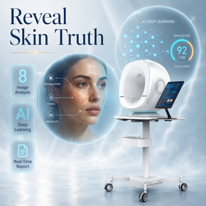
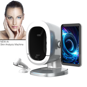
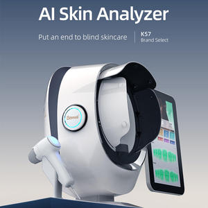

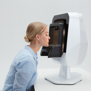
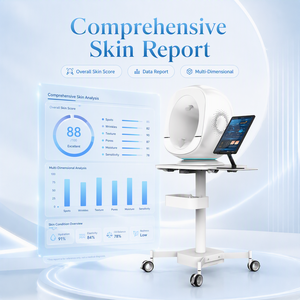
Optical Coherence Tomography (OCT) & 3D Spectral Dermal Imaging for Nigerian Healthcare & Aesthetic Medicine
An authoritative analysis of non-invasive optical biopsies, cross-sectional dermal profiling, and skin phototype IV–VI diagnostic calibrations tailored for West African clinical environments.
1. Executive Summary & Biophysical Foundations of OCT
Optical Coherence Tomography (OCT) represents a monumental paradigm shift in non-invasive clinical dermatology and high-precision aesthetic evaluation. By utilizing low-coherence interferometry, OCT systems project near-infrared light beams into living skin tissues, measuring the optical backscattering delay to construct ultra-high-resolution, micrometer-scale cross-sectional views of dermal architecture. Where traditional visual examinations and surface dermoscopy fail to evaluate sub-surface structural integrity, advanced Optical Coherence Tomography and 3D Multispectral Photometric Stereo Systems provide an non-invasive "in vivo optical biopsy."
For dermatologists, clinical researchers, and aesthetic practitioners in Nigeria—specifically operating in tier-one medical hubs such as Lagos, Abuja, Port Harcourt, and Kano—the demand for non-destructive, quantitative diagnostic equipment has escalated rapidly. The combination of intense ultraviolet irradiation (UV Index frequently exceeding 11+ across Nigeria), high prevalence of post-inflammatory hyperpigmentation (PIH), dermal thinning from uncalibrated cosmetic treatments, and complex skin conditions inherent to Fitzpatrick Phototypes IV, V, and VI requires instrumentation engineered beyond standard European or East Asian baseline presets.
Our proprietary 3D Multispectral Photometric Stereo Engine combined with Swept-Source Optical Coherence Tomography (SS-OCT) algorithms eliminates melanin scattering artifacts inherent to high-melanin cutaneous layers. This allows Nigerian clinicians to accurately quantify sub-epidermal melanin distribution, collagen bundle degradation, vascular erythema, and hair follicle root depth down to 2.5 mm without invasive scalpel biopsies.
2. Optical Physics: Resolving Sub-Surface Layers in Darker Skin Phototypes
The diagnostic challenge in evaluating dark skin phototypes (Fitzpatrick IV–VI) lies in the high concentration of epidermal melanosomes, which absorb and scatter shorter wavelengths of visible light (400–550 nm). Standard clinical skin analyzers often present severe imaging distortion due to surface specular reflections and signal attenuation caused by concentrated epidermal melanin.
Our medical-grade OCT and 9-Spectral AI diagnostic systems circumvent this limitation by deploying narrow-band near-infrared (NIR) wavelengths alongside patented polarising filters and photometric stereo lighting arrays:
- Epidermal-Dermal Junction (EDJ) Visualization: Precisely maps flattening of the rete ridges—a primary biomarker of cutaneous intrinsic aging and structural compromise.
- Melanocyte Depth Profiling: Differentiates between epidermal melasma, dermal melanocytosis, and mixed post-inflammatory hyperpigmentation to guide targeted laser/topical therapy.
- Microvascular Flow Mapping: Detects deep-dermal erythema and teleangiectasia obscured by heavy superficial pigmentation, critical for early rosacea and steroid-induced rosaceiform eruption diagnosis.
- 3D Volumetric Pore & Scar Topography: Measures exact depth (in micrometers) and cubic volume of acne scarring, keloid initiation zones, and enlarged pores for objective pre-and-post procedure verification.
3. Comprehensive Technology Comparison Matrix
To assist healthcare procurement officers, hospital boards, and aesthetic clinic directors in evaluating diagnostic investments, the table below compares standard clinical tools against our advanced Optical Coherence & 3D Multispectral AI Analysis Systems:
| Diagnostic Parameter | Standard Handheld Dermoscopy | 2D RGB Facial Scanners | 3D Multispectral AI & OCT Systems |
|---|---|---|---|
| Penetration Depth | Superficial Stratum Corneum (< 0.1 mm) | Epidermal Surface (0.2 mm) | Full Dermal Layer (1.5 – 2.5 mm) |
| Melanin Depth Segmentation | No (Qualitative Only) | Limited 2D RGB Estimate | Quantitative (Epidermal vs Dermal) |
| Quantitative Scar Volumetrics | None | None | Micron-Level 3D Surface Topography |
| Fitzpatrick IV–VI Calibration | High Refinement Required | Prone to Light Scattering Artifacts | Engineered Dynamic NIR Spectrum |
| Sub-Surface Vascular Clarity | Obscured by Melanin | Poor Visual Distinction | Cross-Polarized Redness Mapping |
| Clinical Publication Readiness | Subjective Observations | Basic Patient CRM Images | Clinical Trial Grade DICOM / Data Export |
4. Localized Clinical Application Scenarios Across Nigeria
Understanding the unique epidemiological and cosmetic landscape of West Africa is essential for delivering meaningful clinical outcomes. Below are key real-world application scenarios where our devices are actively utilized by practitioners in Nigeria:
Post-Chemical Peel & Laser Monitoring: In high-end aesthetic clinics in Victoria Island and Lekki, treating dark skin phototypes with aggressive peels or lasers carries a high risk of post-inflammatory hyperpigmentation (PIH). OCT and multispectral analysis allow aesthetic physicians to measure sub-clinical baseline inflammation and track pigment clearance during recovery, providing mathematical proof of treatment efficacy.
Diagnosis of Bleaching Cream Damage & Steroid Atrophy: Chronic exposure to illicit topical steroids and hydroquinone-rich lightening agents causes profound dermal atrophy, ochronosis, and telangiectasia. Dermal OCT imaging provides non-invasive optical quantification of dermal thinning (epidermal thickness measured in µm), allowing dermatologists in teaching hospitals to document repair trajectory under prescription barrier-restoration protocols.
Keloid & Hypertrophic Scar Mapping: Keloid scar formation occurs with significantly higher frequency in African populations. 3D surface tomography combined with subsurface optical depth analysis assists reconstructive plastic surgeons in evaluating keloid vascularity, fibrous core boundaries, and therapeutic response to intralesional triamcinolone or cryotherapy.
Cosmeceutical Efficacy Validation: As local Nigerian skincare brands expand across Africa, empirical verification of anti-aging, hydration, and hyperpigmentation claims requires objective data. Our 3D scanners provide standardized quantitative metrics accepted by international peer-reviewed journals and regulatory oversight bodies.
5. Localized Development Trends & Market Dynamics in Nigeria (2026–2030)
The Nigerian medical technology landscape is experiencing a fast-paced transformation. Driven by rapid urbanization, expanding middle-class disposable income, and a rising surge in local medical tourism, several dominant market vectors are shaping diagnostic device adoption:
- Transition to Evidence-Based Aesthetic Consultations: Patients in Lagos and Abuja are increasingly sophisticated. The era of subjective visual skin assessment is ending; clients now demand digital proof of skin health before committing to high-value treatment packages (e.g., RF microneedling, laser resurfacing, specialized peels).
- NAFDAC Rigor & Medical Device Standardization: The National Agency for Food and Drug Administration and Control (NAFDAC) is enforcing stricter compliance standards on imported diagnostic equipment. Our devices meet rigorous CE Medical, ISO 13485, and IEC electrical safety certifications, ensuring seamless importation and regulatory clearance.
- Robust Engineering for Tropical & Electrical Environments: Operating medical electronics in West Africa requires resilience against power fluctuations and ambient humidity. Our hardware features wide-voltage power supplies (100V–240V, 50/60Hz compatible with local generator setup), surge protection circuits, and industrial-grade optics sealed against dust ingress.
6. Why Partner With Us: Our Manufacturing & Global OEM Advantage
As a leading global manufacturer specializing in advanced optical dermal analyzers and 3D skin reconstruction engines, we bring clinical credibility and direct B2B manufacturing scale to our Nigerian partners:
Patented 3D Photometric Hardware
Our machines utilize custom optical sensor arrays and multi-spectrum LED configurations, capturing high-frequency micro-relief images without spatial distortion or lens aberration.
AI-Powered Multi-Language Software
Supporting up to 25 operating languages with intuitive cloud integration, automatic report generator algorithms, and customizable branding options for hospitals and private clinic chains.
Dedicated B2B Logistics for Africa
We offer direct air-freight and door-to-door door-to-port logistics support to Murtala Muhammed International Airport (Lagos) and Nnamdi Azikiwe International Airport (Abuja), complete with full documentation.
Frequently Asked Questions for Nigerian Buyers
Addressing key technical, shipping, electrical, and operational inquiries from clinic owners, hospital engineers, and medical supply distributors in Nigeria.
Equip Your Clinic with World-Class Optical Tomography
Empower your clinical practice, elevate patient trust, and obtain factory-direct OEM pricing with full support for Nigerian procurement.
Get Catalog