
Featured Skin Hemoglobin & Multispectral Imaging Systems
Comprehensive analysis of cutting-edge optical analyzers engineered for precise epidermal chromophore mapping, hemoglobin distribution quantification, erythema measurement, and clinical dermatology research in Northern California.
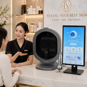
X5PRO 9-Spectral AI Facial Skin Analyzer Machine
Advanced commercial 9-spectrum analyzer specializing in face moisture, deeper skin pore analysis, wrinkles, UV spot visualization, and multi-depth hemoglobin redness mapping. Fully US/EU voltage compatible.
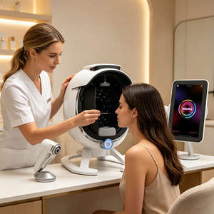
2026 Newest A6 3D Facial Scanner Skin Analyzer
Next-generation 8-spectrum AI facial scanner optimized for deep vascular depth detection, blackheads, pore structural mapping, and baseline hemoglobin concentration profiling for clinical trials.
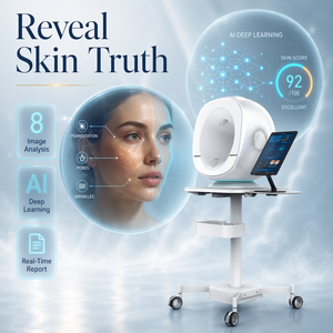
All in One Facial Skin Analyzer Machine 3D Camera
Integrated 4-spectra 8-intelligent image camera system offering 30+ detection parameters. Quantifies sub-surface hemoglobin, vascular inflammation, and dermal topography with high optical resolution.
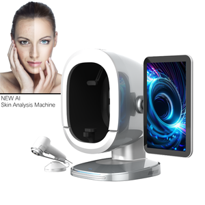
Newest 3D AI Intelligent Skin Analyzer Face Scanner
Smart diagnostic scanner supporting 19 localized operational languages. Features rapid cross-polarized light imaging to separate dermal hemoglobin signals from epidermal melanin artifacts.
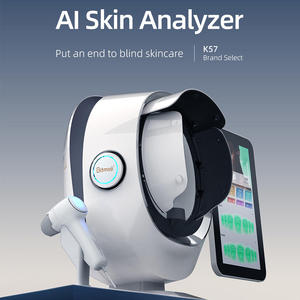
High Quality Portable Facial Analysis Skin Monitor
Compact 3D digital skin analyzer engineered for aesthetic practices, mobile consultations, and cosmetic sales verification. Provides real-time erythema and microvascular mapping.
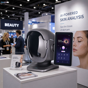
Newest X3-pro-Skin-Analyzer Facial Scanner
High-speed reseller-grade analyzer with 25 pre-installed language packs, direct cloud synchronization, and automated red-blood-cell distribution and vascular lesion assessment.
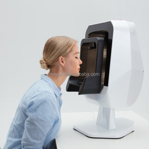
Meicet Mc88 Professional Facial Scanner Machine
Clinical-grade smart beauty machine delivering precise quantitative measurements of hydration, dermal vascularity, inflammation scores, and micro-hemoglobin fluctuations.
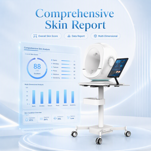
Scientific Research Anti Aging Verification 3D Analyzer
High-precision 4-spectra AI imaging workstation engineered for Contract Research Organizations (CROs) and dermatological studies requiring objective anti-aging and vascular efficacy validation.
Understanding Skin Hemoglobin Quantification in Modern Dermatology
In the rapidly evolving landscape of clinical dermatology, aesthetic medicine, and pharmaceutical formulation testing, non-invasive skin hemoglobin measurement systems have emerged as indispensable tools. Historically, assessing dermal vascularity, inflammation, post-laser erythema, and rosacea relied on subjective visual clinical grading scales—a methodology fraught with inter-observer variability. Today, spectrophotometric chromophore decomposition and 3D multispectral imaging technologies allow researchers and clinicians in the San Francisco Bay Area to map skin hemoglobin concentration with micro-gram per cubic millimeter precision.
Skin color and visual tone are primarily dictated by two biological pigments within the cutaneous architecture: melanin in the basal layer of the epidermis, and hemoglobin (both oxygenated $HbO_2$ and deoxygenated $Hb$) localized within the microvascular network of the papillary and reticular dermis. By utilizing narrow-band spectral illumination and cross-polarized light optics, advanced skin measurement systems isolate the light absorption spectrum of hemoglobin—specifically targeting its absorption peaks at 415 nm, 542 nm, and 577 nm. This optical isolation enables complete separation of vascular redness from melanin pigmentation, giving medical practitioners an unbiased quantitative metric of vascular health, inflammatory reactions, and wound healing kinetics.
Top 10 Skin Hemoglobin System Manufacturers (SF Bay Area Market)
An evaluation based on optical accuracy, multispectral resolution, software compliance (HIPAA/FDA 21 CFR Part 11), local technical support, and clinical research adoption across Northern California.
| Rank & Manufacturer | Primary Optical Technology | Hemoglobin Mapping Depth | Target Sector (SF Bay Area) | Clinical Compliance |
|---|---|---|---|---|
| 1. Miravex Limited (Antera 3D®) | Photometric Stereo + Multispectral | Sub-epidermal (Full Dermis) | Pharma CROs, MedSpas, Clinical Dermatology | Gold Standard |
| 2. Canfield Scientific | RBX® Technology (Red/Brown Separation) | Superficial to Mid Dermis | Academic Medical Centers (UCSF/Stanford) | FDA Registered |
| 3. Courage + Khazaka Electronic | Mexameter® Spectrophotometry | Narrowband Reflectance | Cosmetic Formulation R&D Labs | ISO 13485 |
| 4. Meicet Medical | AI 8-Spectrum Image Processing | Epidermal & Dermal Vascular | Commercial Aesthetic Clinics & Salons | CE / US Compatible |
| 5. QuantifiCare | 3D Stererophotogrammetry | Surface & Erythema Mapping | Plastic Surgery & Injectable Practices | FDA 510(k) Cleared |
| 6. Cortex Technology | DermaSpectrometer / Colorimetry | Point Spectrophotometry | Dermatology Research Labs | Medical Device Class I |
| 7. Bomtech Electronics | Multi-Polarized Facial Scanner | Superficial Redness Mapping | Regional Aesthetic Distributors | CE Certified |
| 8. Observ (Sylton) | Fluorescence & Polarized Light | Vascular Pattern Recognition | Medical Spas & Skincare Centers | Commercial Grade |
| 9. Caliber I.D. | Reflectance Confocal Microscopy | Cellular-Level Capillary Flow | Oncology & Specialized Research | FDA Cleared |
| 10. Spectrumedix Systems | Diffuse Reflectance Spectroscopy | Deep Dermal Microvasculature | Biotech Therapeutics (South SF) | Research Use Only (RUO) |
In-Depth Analysis of Regional Market Leaders
1. Miravex Limited (Antera 3D® Platform): Renowned globally and widely adopted throughout Northern California’s top medical aesthetic centers and biopharmaceutical research institutes, Miravex sets the technical benchmark with its patented Antera 3D® skin imaging technology. Unlike traditional flat 2D cameras, the system utilizes photometric stereo reconstruction combined with multispectral light-emitting diodes (LEDs) spanning the visible spectrum. By capturing skin illumination from multiple precise directions, the system extracts the true 3D topography of the skin surface while simultaneously calculating the absorption coefficients of hemoglobin and melanin. This allows clinicians to quantify total hemoglobin concentration, assess skin redness, track vascular healing post-IPL or pulse-dye laser (PDL) treatments, and measure inflammation reduction in clinical drug trials with absolute mathematical reproducibility.
2. Canfield Scientific: Headquartered nationally with strong regional service infrastructure in California, Canfield’s RBX® technology separates red and brown skin components to visual aesthetic consultation rooms. While highly effective for patient communication in plastic surgery and aesthetic dermatology, its reliance on fixed high-resolution studio hardware makes it less portable compared to hand-held spectrophotometric devices.
3. Courage + Khazaka Electronic: A traditional pillar in cosmetic science laboratories, their Mexameter® probe measures narrow-band reflectance at 568 nm (corresponding to an absorption peak of hemoglobin) and 880 nm. It remains a standard point-measurement tool for cosmetic chemists in the Bay Area evaluating anti-inflammatory botanical extracts.
Enterprise Optical & Software Architecture
Our featured hemoglobin analysis systems incorporate proprietary photometric stereo imaging algorithms and multi-spectral extraction engines designed to deliver clinical-grade validation for pharmaceutical manufacturers, CROs, and high-volume medical spas.
How Multispectral Hemoglobin Mapping Works
1. Spectral Illumination
Illuminating the tissue with specific narrow-band optical wavelengths allows the optical sensor to target the unique absorption bands of oxyhemoglobin ($HbO_2$) and deoxyhemoglobin ($Hb$) while excluding scattering noise.
2. Photometric Stereo 3D
By sequential illumination from multiple elevation angles, the system reconstructs the surface normal vectors of the skin, decoupling surface reflection (glare) from diffuse sub-surface dermal reflectance.
3. Quantitative Hemoglobin Concentration
Mathematical matrix algorithms transform multi-spectral RGB data into true physiological concentration maps ($C_{Hb}$ in $mg/cm^3$), rendering objective numerical values for vascular lesions and erythema.
Localized Application Scenarios across the San Francisco Bay Area
Northern California represents a unique synthesis of biomedical innovation, clinical academic research, and high-end aesthetic medicine. Skin hemoglobin measurement systems play a pivotal role across three major regional hubs.
A. South San Francisco Biotech Corridor & CRO Clinical Trials
Home to the world's largest concentration of biotechnology firms, South San Francisco clinical contract research organizations (CROs) utilize quantitative hemoglobin imaging to validate topical dermatological therapeutics. In phase II and phase III clinical trials for psoriasis, eczema, rosacea, and post-procedural anti-inflammatory topicals, objective measurements of skin redness are legally required for FDA submission dossiers. Portable multispectral analyzers allow clinical trial coordinators to maintain zero intra-operator error across multi-center studies.
B. Premium Aesthetic Clinics in Downtown SF, Palo Alto & Marin County
High-end aesthetic practices in San Francisco's Union Square, Palo Alto, and Walnut Creek cater to tech executives and discerning clientele seeking quantifiable outcomes. Integrating 3D skin analyzers for pre-treatment consultation allows practitioners to demonstrate hidden sub-surface microvascular damage, spider veins, and telangiectasia. Following Intense Pulsed Light (IPL), Vascular Lasers (Pulsed Dye / 1064nm Nd:YAG), or Microneedling RF procedures, baseline vs. post-treatment hemoglobin heatmaps confirm treatment efficacy, driving patient retention and treatment plan compliance.
C. Academic Research at UCSF & Stanford University Medical Centers
Academic dermatologists at UCSF Department of Dermatology and Stanford Medicine use advanced 3D photometric skin scanners for non-invasive longitudinal monitoring of cutaneous vascular disorders, skin graft perfusion assessment, and microvascular changes associated with autoimmune conditions such as systemic sclerosis and lupus erythematosus.
San Francisco Regional Trends in Optical Skin Diagnostics
1. AI-Driven Microvascular Diagnostics: The integration of deep learning algorithms into skin analyzers allows automatic categorization of facial redness into distinct pathologies: diffuse erythema, rosacea subtype 1/2, acne-induced post-inflammatory erythema (PIE), or broken capillaries. Silicon Valley AI research groups are partnering with hardware suppliers to train neural networks on multi-ethnic datasets.
2. Multi-Ethnic & Darker Skin Tone Calibration: San Francisco features an exceptionally diverse demographic composition. Traditional RGB camera systems often fail to accurately separate melanin from hemoglobin in Fitzpatrick Skin Types IV through VI because high epidermal melanin absorbs visible light intensely. Modern narrow-band cross-polarized spectro-imaging systems filter out surface melanin scattering, ensuring equitable diagnostic accuracy for all patient demographics across Northern California.
3. Cloud-Connected Tele-Dermatology Networks: With remote healthcare adoption booming across the Bay Area, clinics are deploying compact 3D skin analyzers capable of instant HIPAA-compliant cloud syncing. Practitioners can track patient vascular healing remotely, export quantitative PDF reports into Electronic Health Record (EHR) platforms, and streamline multi-clinic management.
Frequently Asked Questions by San Francisco Buyers
Essential technical, regulatory, and ROI insights for Bay Area dermatologists, plastic surgeons, CRO managers, and aesthetic practice directors.
Q: How does multispectral hemoglobin mapping differ from standard RGB skin camera images?
Standard RGB cameras capture broad red, green, and blue light channels that overlap significantly, causing superficial melanin pigmentation and light reflection to distort redness measurements. Multispectral skin hemoglobin analyzers utilize cross-polarized illumination and precise narrow-band optical filters tuned to the absorption spectrum of hemoglobin (542nm and 577nm). This optical isolation mathematically removes epidermal melanin interference, providing true concentration maps of sub-surface vascularity.
Q: Are these systems compliant with California Medical Board & HIPAA privacy standards?
Yes. Clinical-grade systems designed for US commercial deployment incorporate AES 256-bit encryption for stored patient images and diagnostic data. Software platforms allow anonymized data exports compliant with HIPAA regulations, making them safe for clinical research, cloud backups, and digital patient consultations across California health networks.
Q: What is the expected ROI for a San Francisco MedSpa or aesthetic dermatology practice?
Bay Area practices typically report full device ROI within 3 to 6 months. By presenting objective baseline hemoglobin and vascular maps during patient consultations, practices experience an estimated 35% increase in conversion rates for vascular laser packages (such as IPL, VBeam, or Nd:YAG) and skincare regimens. Post-treatment scans confirm tangible results, boosting patient retention and package renewals.
Q: Can these systems measure both skin texture/wrinkles and hemoglobin simultaneously?
Yes. Systems utilizing 3D Photometric Stereo technology (such as the Antera 3D® and featured 8-spectrum AI scanners) capture mathematical 3D surface elevation alongside spectral color data in a single acquisition under 2 seconds. This enables simultaneous quantification of skin roughness ($Ra$, $Rq$), wrinkle depth, pore volume, melanin index, and vascular concentration.
Q: What calibration protocols are required for high-throughput CRO clinical trials?
For clinical research applications in South San Francisco, hardware systems include standardized optical calibration charts (color and reflectance standards). Daily calibration takes less than 30 seconds, ensuring zero drift in illumination intensity or sensor response over multi-month clinical study timelines.
Q: What technical support and training options are available in Northern California?
Leading suppliers offer direct local on-site installation, staff clinical training, software integration assistance, and rapid-replacement warranty programs within the San Francisco Bay Area, Silicon Valley, and Greater Sacramento regions.
Upgrade Your Practice with Clinical 3D Hemoglobin Analysis
Connect with our skin diagnostic technology specialists to request technical datasheets, request an on-site demonstration in the San Francisco Bay Area, or obtain competitive commercial quotations.